 Movie
Movie Controller

[English] 日本語
 Yorodumi
- PDB-8tdr: Crystal structure of the methyltransferase domain of DNMT3A homot... -
+ Open data

- Basic information
| Entry | Database: PDB / ID: 8tdr | ||||||
|---|---|---|---|---|---|---|---|
| Title | Crystal structure of the methyltransferase domain of DNMT3A homotetramer | ||||||
 Components Components | DNA (cytosine-5)-methyltransferase 3A | ||||||
 Keywords Keywords | TRANSFERASE / DNA Methyltransferase | ||||||
| Function / homology |  Function and homology information Function and homology informationpositive regulation of cellular response to hypoxia / transposable element silencing by piRNA-mediated DNA methylation / protein-cysteine methyltransferase activity / regulatory ncRNA-mediated heterochromatin formation / unmethylated CpG binding / cellular response to bisphenol A / DNA (cytosine-5-)-methyltransferase / DNA (cytosine-5-)-methyltransferase activity / autosome genomic imprinting / SUMOylation of DNA methylation proteins ...positive regulation of cellular response to hypoxia / transposable element silencing by piRNA-mediated DNA methylation / protein-cysteine methyltransferase activity / regulatory ncRNA-mediated heterochromatin formation / unmethylated CpG binding / cellular response to bisphenol A / DNA (cytosine-5-)-methyltransferase / DNA (cytosine-5-)-methyltransferase activity / autosome genomic imprinting / SUMOylation of DNA methylation proteins / XY body / response to vitamin A / response to ionizing radiation / DNA methylation-dependent constitutive heterochromatin formation / negative regulation of gene expression via chromosomal CpG island methylation / hepatocyte apoptotic process / lncRNA binding / cellular response to ethanol / chromosome, centromeric region / catalytic complex / heterochromatin / Transferases; Transferring one-carbon groups; Methyltransferases / DNA methylation / PRC2 methylates histones and DNA / post-embryonic development / Defective pyroptosis / response to cocaine / cellular response to amino acid stimulus / Regulation of endogenous retroelements by Piwi-interacting RNAs (piRNAs) / euchromatin / response to lead ion / response to toxic substance / nuclear matrix / RMTs methylate histone arginines / neuron differentiation / transcription corepressor activity / response to estradiol / spermatogenesis / methylation / cellular response to hypoxia / RNA polymerase II-specific DNA-binding transcription factor binding / RNA polymerase II cis-regulatory region sequence-specific DNA binding / response to xenobiotic stimulus / negative regulation of DNA-templated transcription / chromatin binding / negative regulation of transcription by RNA polymerase II / DNA binding / zinc ion binding / nucleoplasm / identical protein binding / nucleus / cytoplasm Similarity search - Function | ||||||
| Biological species |  Homo sapiens (human) Homo sapiens (human) | ||||||
| Method |  X-RAY DIFFRACTION / SYNCHROTRON / MOLECULAR REPLACEMENT / Resolution: 3.32 Å X-RAY DIFFRACTION / SYNCHROTRON / MOLECULAR REPLACEMENT / Resolution: 3.32 Å | ||||||
 Authors Authors | Lu, J.W. / Song, J.K. | ||||||
| Funding support |  United States, 1items United States, 1items
| ||||||
 Citation Citation | Journal: Nat Commun / Year: 2024 Title: Structure-guided functional suppression of AML-associated DNMT3A hotspot mutations. Authors: Lu, J. / Guo, Y. / Yin, J. / Chen, J. / Wang, Y. / Wang, G.G. / Song, J. | ||||||
| History |
|
- Structure visualization
| Structure viewer | Molecule: MolmilJmol/JSmol |
|---|
- Downloads & links
-Download
| PDBx/mmCIF format | 8tdr.cif.gz | 726.1 KB | Display | PDBx/mmCIF format |
|---|---|---|---|---|
| PDB format | pdb8tdr.ent.gz | 598 KB | Display | PDB format |
| PDBx/mmJSON format | 8tdr.json.gz | Tree view | PDBx/mmJSON format | |
| Others |  Other downloads Other downloads |
-Validation report
| Summary document | 8tdr_validation.pdf.gz | 1.3 MB | Display | wwPDB validaton report |
|---|---|---|---|---|
| Full document | 8tdr_full_validation.pdf.gz | 1.4 MB | Display | |
| Data in XML | 8tdr_validation.xml.gz | 67.7 KB | Display | |
| Data in CIF | 8tdr_validation.cif.gz | 85.8 KB | Display | |
| Arichive directory | https://data.pdbj.org/pub/pdb/validation_reports/td/8tdrftp://data.pdbj.org/pub/pdb/validation_reports/td/8tdr | HTTPS FTP |
-Related structure data
| Related structure data | 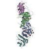 8te1C 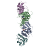 8te3C  8te4C 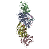 5yx2S S: Starting model for refinement C: citing same article ( |
|---|---|
| Similar structure data |
-Links
 PDBj
PDBj

- Assembly
| Deposited unit | 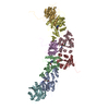
| ||||||||
|---|---|---|---|---|---|---|---|---|---|
| 1 | 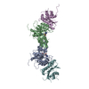
| ||||||||
| 2 | 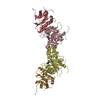
| ||||||||
| Unit cell |
|
-Components
| #1: Protein | Mass: 32859.844 Da / Num. of mol.: 8 / Fragment: methyltransferase domain (UNP residues 628-912) Source method: isolated from a genetically manipulated source Source: (gene. exp.) Homo sapiens (human) / Gene: DNMT3A / Production host:  References: UniProt: Q9Y6K1, DNA (cytosine-5-)-methyltransferase, Transferases; Transferring one-carbon groups; Methyltransferases #2: Chemical | ChemComp-SAH /   Mass: 384.411 Da / Num. of mol.: 4 / Source method: obtained synthetically / Formula: C14H20N6O5S / Feature type: SUBJECT OF INVESTIGATION Mass: 384.411 Da / Num. of mol.: 4 / Source method: obtained synthetically / Formula: C14H20N6O5S / Feature type: SUBJECT OF INVESTIGATIONHas ligand of interest | Y | |
|---|
-Experimental details
-Experiment
| Experiment | Method: X-RAY DIFFRACTION / Number of used crystals: 1 |
|---|
- Sample preparation
| Crystal | Density Matthews: 3.82 Å3/Da / Density % sol: 67.81 % |
|---|---|
| Crystal grow | Temperature: 277 K / Method: vapor diffusion, sitting drop / Details: 0.2-0.4 M potassium sodium tartrate |
-Data collection
| Diffraction | Mean temperature: 100 K / Serial crystal experiment: N | |||||||||||||||||||||||||||||||||||||||||||||||||||||||||||||||||||||||||||||||||||||||||||||||||||
|---|---|---|---|---|---|---|---|---|---|---|---|---|---|---|---|---|---|---|---|---|---|---|---|---|---|---|---|---|---|---|---|---|---|---|---|---|---|---|---|---|---|---|---|---|---|---|---|---|---|---|---|---|---|---|---|---|---|---|---|---|---|---|---|---|---|---|---|---|---|---|---|---|---|---|---|---|---|---|---|---|---|---|---|---|---|---|---|---|---|---|---|---|---|---|---|---|---|---|---|---|
| Diffraction source | Source: SYNCHROTRON / Site: ALS / Beamline: 5.0.2 / Wavelength: 1 Å | |||||||||||||||||||||||||||||||||||||||||||||||||||||||||||||||||||||||||||||||||||||||||||||||||||
| Detector | Type: DECTRIS PILATUS3 6M / Detector: PIXEL / Date: Feb 7, 2020 | |||||||||||||||||||||||||||||||||||||||||||||||||||||||||||||||||||||||||||||||||||||||||||||||||||
| Radiation | Protocol: SINGLE WAVELENGTH / Monochromatic (M) / Laue (L): M / Scattering type: x-ray | |||||||||||||||||||||||||||||||||||||||||||||||||||||||||||||||||||||||||||||||||||||||||||||||||||
| Radiation wavelength | Wavelength: 1 Å / Relative weight: 1 | |||||||||||||||||||||||||||||||||||||||||||||||||||||||||||||||||||||||||||||||||||||||||||||||||||
| Reflection | Resolution: 3.32→50 Å / Num. obs: 57297 / % possible obs: 100 % / Redundancy: 3.9 % / Biso Wilson estimate: 84.77 Å2 / Rmerge(I) obs: 0.22 / Rpim(I) all: 0.127 / Rrim(I) all: 0.254 / Χ2: 0.583 / Net I/σ(I): 3.3 / Num. measured all: 225061 | |||||||||||||||||||||||||||||||||||||||||||||||||||||||||||||||||||||||||||||||||||||||||||||||||||
| Reflection shell | Diffraction-ID: 1
|
- Processing
| Software |
| |||||||||||||||||||||||||||||||||||||||||||||||||||||||||||||||||||||||||||||||||||||||||||||||||||||||||
|---|---|---|---|---|---|---|---|---|---|---|---|---|---|---|---|---|---|---|---|---|---|---|---|---|---|---|---|---|---|---|---|---|---|---|---|---|---|---|---|---|---|---|---|---|---|---|---|---|---|---|---|---|---|---|---|---|---|---|---|---|---|---|---|---|---|---|---|---|---|---|---|---|---|---|---|---|---|---|---|---|---|---|---|---|---|---|---|---|---|---|---|---|---|---|---|---|---|---|---|---|---|---|---|---|---|---|
| Refinement | Method to determine structure: MOLECULAR REPLACEMENT Starting model: PDB entry 5YX2 Resolution: 3.32→46.29 Å / SU ML: 0.46 / Cross valid method: THROUGHOUT / σ(F): 1.96 / Phase error: 27.64 / Stereochemistry target values: ML
| |||||||||||||||||||||||||||||||||||||||||||||||||||||||||||||||||||||||||||||||||||||||||||||||||||||||||
| Solvent computation | Shrinkage radii: 0.9 Å / VDW probe radii: 1.1 Å / Solvent model: FLAT BULK SOLVENT MODEL | |||||||||||||||||||||||||||||||||||||||||||||||||||||||||||||||||||||||||||||||||||||||||||||||||||||||||
| Displacement parameters | Biso max: 193.69 Å2 / Biso mean: 81.129 Å2 / Biso min: 30 Å2 | |||||||||||||||||||||||||||||||||||||||||||||||||||||||||||||||||||||||||||||||||||||||||||||||||||||||||
| Refinement step | Cycle: final / Resolution: 3.32→46.29 Å
| |||||||||||||||||||||||||||||||||||||||||||||||||||||||||||||||||||||||||||||||||||||||||||||||||||||||||
| LS refinement shell | Refine-ID: X-RAY DIFFRACTION / Rfactor Rfree error: 0 / Total num. of bins used: 14
| |||||||||||||||||||||||||||||||||||||||||||||||||||||||||||||||||||||||||||||||||||||||||||||||||||||||||
| Refinement TLS params. | Method: refined / Origin x: 36.4286 Å / Origin y: -51.9343 Å / Origin z: 34.8961 Å
| |||||||||||||||||||||||||||||||||||||||||||||||||||||||||||||||||||||||||||||||||||||||||||||||||||||||||
| Refinement TLS group |
|
