 Movie
Movie Controller

[English] 日本語
 Yorodumi
- PDB-8tcl: Crystal Structure of modified HIV reverse transcriptase p51 domai... -
+ Open data

- Basic information
| Entry | Database: PDB / ID: 8tcl | |||||||||
|---|---|---|---|---|---|---|---|---|---|---|
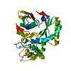
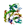
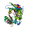
| Title | Crystal Structure of modified HIV reverse transcriptase p51 domain (FPC2) with picrate bound | |||||||||
 Components Components | p51 subunit | |||||||||
 Keywords Keywords | DNA BINDING PROTEIN / P51 subunit / HIV / AIDS / transferase / picric acid | |||||||||
| Function / homology | PICRIC ACID Function and homology information Function and homology information | |||||||||
| Biological species | HIV whole-genome vector AA1305#18 (others) | |||||||||
| Method |  X-RAY DIFFRACTION / MOLECULAR REPLACEMENT / Resolution: 1.95 Å X-RAY DIFFRACTION / MOLECULAR REPLACEMENT / Resolution: 1.95 Å | |||||||||
 Authors Authors | Pedersen, L.C. / London, R.E. | |||||||||
| Funding support |  United States, 2items United States, 2items
| |||||||||
 Citation Citation | Journal: Biomolecules / Year: 2023 Title: Targeting the Structural Maturation Pathway of HIV-1 Reverse Transcriptase. Authors: Kirby, T.W. / Gabel, S.A. / DeRose, E.F. / Perera, L. / Krahn, J.M. / Pedersen, L.C. / London, R.E. | |||||||||
| History |
|
- Structure visualization
| Structure viewer | Molecule: MolmilJmol/JSmol |
|---|
- Downloads & links
-Download
| PDBx/mmCIF format | 8tcl.cif.gz | 145.3 KB | Display | PDBx/mmCIF format |
|---|---|---|---|---|
| PDB format | pdb8tcl.ent.gz | 112 KB | Display | PDB format |
| PDBx/mmJSON format | 8tcl.json.gz | Tree view | PDBx/mmJSON format | |
| Others |  Other downloads Other downloads |
-Validation report
| Arichive directory | https://data.pdbj.org/pub/pdb/validation_reports/tc/8tclftp://data.pdbj.org/pub/pdb/validation_reports/tc/8tcl | HTTPS FTP |
|---|
-Related structure data
-Links
 PDBj
PDBj
- Assembly
| Deposited unit | 
| |||||||||
|---|---|---|---|---|---|---|---|---|---|---|
| 1 |
| |||||||||
| Unit cell |
| |||||||||
| Components on special symmetry positions |
|
-Components
| #1: Protein | Mass: 39805.246 Da / Num. of mol.: 1 / Mutation: E203S Source method: isolated from a genetically manipulated source Source: (gene. exp.) HIV whole-genome vector AA1305#18 (others) Production host:  | ||||||
|---|---|---|---|---|---|---|---|
| #2: Chemical |   Mass: 229.104 Da / Num. of mol.: 3 / Source method: obtained synthetically / Formula: C6H3N3O7 / Feature type: SUBJECT OF INVESTIGATION Mass: 229.104 Da / Num. of mol.: 3 / Source method: obtained synthetically / Formula: C6H3N3O7 / Feature type: SUBJECT OF INVESTIGATION#3: Water | ChemComp-HOH / |  Mass: 18.015 Da / Num. of mol.: 147 / Source method: isolated from a natural source / Formula: H2O Mass: 18.015 Da / Num. of mol.: 147 / Source method: isolated from a natural source / Formula: H2OHas ligand of interest | Y | Has protein modification | N | |
-Experimental details
-Experiment
| Experiment | Method: X-RAY DIFFRACTION / Number of used crystals: 1 |
|---|
- Sample preparation
| Crystal | Density Matthews: 2.53 Å3/Da / Density % sol: 51.35 % |
|---|---|
| Crystal grow | Temperature: 277 K / Method: vapor diffusion, sitting drop Details: protein: 25mg/ml 10mM Tris pH 7.4, 40mM NaCl, 1mM TCEP, 0.25mM azide, 10mM picric acid ML: 20%PEG3350, 0.2M trisodium citrate |
-Data collection
| Diffraction | Mean temperature: 100 K / Serial crystal experiment: N |
|---|---|
| Diffraction source | Source: ROTATING ANODE / Type: RIGAKU MICROMAX-007 HF / Wavelength: 1.514 Å |
| Detector | Type: RIGAKU SATURN 944+ / Detector: CCD / Date: Sep 19, 2019 / Details: VarimaxHF |
| Radiation | Protocol: SINGLE WAVELENGTH / Monochromatic (M) / Laue (L): M / Scattering type: x-ray |
| Radiation wavelength | Wavelength: 1.514 Å / Relative weight: 1 |
| Reflection | Resolution: 1.95→50 Å / Num. obs: 28616 / % possible obs: 96.1 % / Redundancy: 4.2 % / Rpim(I) all: 0.024 / Rrim(I) all: 0.054 / Rsym value: 0.048 / Net I/σ(I): 10.7 |
| Reflection shell | Resolution: 1.95→1.98 Å / Num. unique obs: 1214 / CC1/2: 0.862 / Rpim(I) all: 9.268 / Rrim(I) all: 0.473 / Rsym value: 0.386 / % possible all: 84.1 |
- Processing
| Software |
| |||||||||||||||||||||||||||||||||||||||||||||||||||||||||||||||||||||||||||||||||||||||||||||||||||||||||||||||||||||||||||||
|---|---|---|---|---|---|---|---|---|---|---|---|---|---|---|---|---|---|---|---|---|---|---|---|---|---|---|---|---|---|---|---|---|---|---|---|---|---|---|---|---|---|---|---|---|---|---|---|---|---|---|---|---|---|---|---|---|---|---|---|---|---|---|---|---|---|---|---|---|---|---|---|---|---|---|---|---|---|---|---|---|---|---|---|---|---|---|---|---|---|---|---|---|---|---|---|---|---|---|---|---|---|---|---|---|---|---|---|---|---|---|---|---|---|---|---|---|---|---|---|---|---|---|---|---|---|---|
| Refinement | Method to determine structure: MOLECULAR REPLACEMENT / Resolution: 1.95→21.62 Å / SU ML: 0.18 / Cross valid method: FREE R-VALUE / σ(F): 1.33 / Phase error: 26.04 / Stereochemistry target values: ML
| |||||||||||||||||||||||||||||||||||||||||||||||||||||||||||||||||||||||||||||||||||||||||||||||||||||||||||||||||||||||||||||
| Solvent computation | Shrinkage radii: 0.9 Å / VDW probe radii: 1.11 Å / Solvent model: FLAT BULK SOLVENT MODEL | |||||||||||||||||||||||||||||||||||||||||||||||||||||||||||||||||||||||||||||||||||||||||||||||||||||||||||||||||||||||||||||
| Refinement step | Cycle: LAST / Resolution: 1.95→21.62 Å
| |||||||||||||||||||||||||||||||||||||||||||||||||||||||||||||||||||||||||||||||||||||||||||||||||||||||||||||||||||||||||||||
| Refine LS restraints |
| |||||||||||||||||||||||||||||||||||||||||||||||||||||||||||||||||||||||||||||||||||||||||||||||||||||||||||||||||||||||||||||
| LS refinement shell |
| |||||||||||||||||||||||||||||||||||||||||||||||||||||||||||||||||||||||||||||||||||||||||||||||||||||||||||||||||||||||||||||
| Refinement TLS params. | Method: refined / Refine-ID: X-RAY DIFFRACTION
| |||||||||||||||||||||||||||||||||||||||||||||||||||||||||||||||||||||||||||||||||||||||||||||||||||||||||||||||||||||||||||||
| Refinement TLS group |
|
