[English] 日本語
Yorodumi
- PDB-8t8a: Structure of arginine oxidase from Pseudomonas sp. TRU 7192 -

+
Open data


ID or keywords:

Loading...

-
Basic information

Entry
Database: PDB / ID: 8t8a
TitleStructure of arginine oxidase from Pseudomonas sp. TRU 7192
ComponentsAmine oxidoreductase
KeywordsOXIDOREDUCTASE / oxidase / arginine oxidase
Function / homologytryptophan 2-monooxygenase / auxin biosynthetic process / : / Amine oxidase / Flavin containing amine oxidoreductase / monooxygenase activity / FAD/NAD(P)-binding domain superfamily / FLAVIN-ADENINE DINUCLEOTIDE / Tryptophan 2-monooxygenase
Function and homology information
Biological speciesPseudomonas sp. (bacteria)
MethodX-RAY DIFFRACTION / SYNCHROTRON / MOLECULAR REPLACEMENT / Resolution: 3.4 Å
AuthorsTakahashi, K. / Yamaguchi, H. / Tatsumi, M. / Sugiki, M.
Funding support1items
OrganizationGrant numberCountry
Not funded
Citation
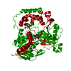
Journal: J Biochem / Year: 2025
Title: Open and closed structures of L-arginine oxidase by cryo-electron microscopy and X-ray crystallography.
Authors: Hiroki Yamaguchi / Kazutoshi Takahashi / Nobutaka Numoto / Hiroshi Suzuki / Moemi Tatsumi / Akiko Kamegawa / Kouki Nishikawa / Yasuhisa Asano / Toshimi Mizukoshi / Hiroshi Miyano / Yoshinori ...Authors: Hiroki Yamaguchi / Kazutoshi Takahashi / Nobutaka Numoto / Hiroshi Suzuki / Moemi Tatsumi / Akiko Kamegawa / Kouki Nishikawa / Yasuhisa Asano / Toshimi Mizukoshi / Hiroshi Miyano / Yoshinori Fujiyoshi / Masayuki Sugiki /
Abstract: L-arginine oxidase (AROD, EC 1.4.3.25) is an oxidoreductase that catalyses the deamination of L-arginine, with flavin adenine dinucleotide (FAD) as a cofactor. Recently identified AROD from ...L-arginine oxidase (AROD, EC 1.4.3.25) is an oxidoreductase that catalyses the deamination of L-arginine, with flavin adenine dinucleotide (FAD) as a cofactor. Recently identified AROD from Pseudomonas sp. TPU 7192 (PT-AROD) demonstrates high selectivity for L-arginine. This enzyme is useful for accurate assays of L-arginine in biological samples. The structural characteristics of the FAD-dependent AROD, however, remain unknown. Here, we report the structure of PT-AROD at a resolution of 2.3 Å by cryo-electron microscopy. PT-AROD adopts an octameric structure with D4 symmetry, which is consistent with its molecular weight in solution, estimated by mass photometry. Comparative analysis of this structure with that determined using X-ray crystallography reveals open and closed forms of the lid-like loop at the entrance to the substrate pocket. Furthermore, mutation of Glu493, located at the substrate binding site, diminishes substrate selectivity, suggesting that this residue contributes significantly to the high selectivity of PT-AROD.
History
DepositionJun 22, 2023Deposition site: RCSB / Processing site: RCSB
Revision 1.0Jun 26, 2024Provider: repository / Type: Initial release
Revision 1.1Oct 23, 2024Group: Structure summary / Category: pdbx_entry_details / pdbx_modification_feature / Item: _pdbx_entry_details.has_protein_modification
Revision 1.2Jan 8, 2025Group: Database references / Category: citation / citation_author
Item: _citation.country / _citation.journal_abbrev ..._citation.country / _citation.journal_abbrev / _citation.journal_id_CSD / _citation.journal_id_ISSN / _citation.pdbx_database_id_DOI / _citation.pdbx_database_id_PubMed / _citation.title / _citation.year
Revision 1.3Jan 15, 2025Group: Database references / Category: citation
Item: _citation.journal_volume / _citation.page_first ..._citation.journal_volume / _citation.page_first / _citation.page_last / _citation.year

-
Structure visualization

Structure viewerMolecule:
MolmilJmol/JSmol

Downloads & links

-
Assembly

Deposited unit
A: Amine oxidoreductase
B: Amine oxidoreductase
C: Amine oxidoreductase
D: Amine oxidoreductase
hetero molecules


Theoretical massNumber of molelcules
Total (without water)272,8358
Polymers269,6934
Non-polymers3,1424
Water2,072115
1
A: Amine oxidoreductase
B: Amine oxidoreductase
C: Amine oxidoreductase
D: Amine oxidoreductase
hetero molecules

A: Amine oxidoreductase
B: Amine oxidoreductase
C: Amine oxidoreductase
D: Amine oxidoreductase
hetero molecules


Theoretical massNumber of molelcules
Total (without water)545,67016
Polymers539,3868
Non-polymers6,2848
Water1448
TypeNameSymmetry operationNumber
identity operation1_555x,y,z1
crystal symmetry operation4_565x,-y+1,-z1
Buried area41660 Å2
ΔGint-204 kcal/mol
Surface area160300 Å2
MethodPISA
Unit cell
Length a, b, c (Å)168.274, 200.219, 168.542
Angle α, β, γ (deg.)90.00, 90.00, 90.00
Int Tables number20
Space group name H-MC2221

-
Components

#1: Protein
Amine oxidoreductase


Mass: 67423.188 Da / Num. of mol.: 4
Source method: isolated from a genetically manipulated source
Source: (gene. exp.) Pseudomonas sp. (bacteria) / Strain: TPU 7192 / Gene: AYO28_23740 / Production host: Escherichia coli (E. coli) / References: UniProt: A0A177SH44
#2: Chemical
ChemComp-FAD / FLAVIN-ADENINE DINUCLEOTIDE


Mass: 785.550 Da / Num. of mol.: 4 / Source method: isolated from a natural source / Formula: C27H33N9O15P2 / Feature type: SUBJECT OF INVESTIGATION / Comment: FAD*YM
#3: Water ChemComp-HOH / water


Mass: 18.015 Da / Num. of mol.: 115 / Source method: isolated from a natural source / Formula: H2O
Has ligand of interestY
Has protein modificationY

-
Experimental details

-
Experiment

ExperimentMethod: X-RAY DIFFRACTION / Number of used crystals: 1

-
Sample preparation

CrystalDensity Matthews: 2.7 Å3/Da / Density % sol: 54.45 %
Crystal growTemperature: 298 K / Method: vapor diffusion, hanging drop / Details: 20% PEG3350, 0.2M Potassium citrate

-
Data collection

DiffractionMean temperature: 100 K / Serial crystal experiment: N
Diffraction sourceSource: SYNCHROTRON / Site: Photon Factory / Beamline: BL-5A / Wavelength: 1 Å
DetectorType: DECTRIS PILATUS3 S 6M / Detector: PIXEL / Date: Jun 29, 2018
RadiationProtocol: SINGLE WAVELENGTH / Monochromatic (M) / Laue (L): M / Scattering type: x-ray
Radiation wavelengthWavelength: 1 Å / Relative weight: 1
ReflectionResolution: 3.4→49.23 Å / Num. obs: 39436 / % possible obs: 100 % / Redundancy: 6.7 % / Rmerge(I) obs: 0.346 / Rpim(I) all: 0.144 / Net I/σ(I): 4.6
Reflection shellResolution: 3.4→3.47 Å / Rmerge(I) obs: 0.977 / Mean I/σ(I) obs: 2.1 / Num. unique obs: 4532 / Rpim(I) all: 0.401 / % possible all: 100

-
Processing

Software
NameVersionClassification
REFMAC5.8.0258refinement
XDSdata reduction
Aimlessdata scaling
PHASERphasing
RefinementMethod to determine structure: MOLECULAR REPLACEMENT / Resolution: 3.4→49.04 Å / Cor.coef. Fo:Fc: 0.937 / Cor.coef. Fo:Fc free: 0.845 / SU B: 50.64 / SU ML: 0.749 / Cross valid method: THROUGHOUT / ESU R Free: 0.748 / Stereochemistry target values: MAXIMUM LIKELIHOOD / Details: HYDROGENS HAVE BEEN ADDED IN THE RIDING POSITIONS
RfactorNum. reflection% reflectionSelection details
Rfree0.31298 1970 5 %RANDOM
Rwork0.21198 ---
obs0.21698 37466 99.92 %-
Solvent computationIon probe radii: 0.8 Å / Shrinkage radii: 0.8 Å / VDW probe radii: 1.2 Å / Solvent model: MASK
Displacement parametersBiso mean: 82.559 Å2
Baniso -1Baniso -2Baniso -3
1--13.5 Å2-0 Å20 Å2
2--3.51 Å2-0 Å2
3---9.99 Å2
Refinement stepCycle: 1 / Resolution: 3.4→49.04 Å
ProteinNucleic acidLigandSolventTotal
Num. atoms17447 0 212 115 17774
Refine LS restraints
Refine-IDTypeDev idealDev ideal targetNumber
X-RAY DIFFRACTIONr_bond_refined_d0.0040.01318252
X-RAY DIFFRACTIONr_bond_other_d0.0010.01715655
X-RAY DIFFRACTIONr_angle_refined_deg1.4211.64524984
X-RAY DIFFRACTIONr_angle_other_deg1.1561.57136127
X-RAY DIFFRACTIONr_dihedral_angle_1_deg7.3552327
X-RAY DIFFRACTIONr_dihedral_angle_2_deg36.43222.399917
X-RAY DIFFRACTIONr_dihedral_angle_3_deg22.65152491
X-RAY DIFFRACTIONr_dihedral_angle_4_deg18.73915105
X-RAY DIFFRACTIONr_chiral_restr0.0490.22344
X-RAY DIFFRACTIONr_gen_planes_refined0.0050.0221148
X-RAY DIFFRACTIONr_gen_planes_other0.0020.023971
X-RAY DIFFRACTIONr_nbd_refined
X-RAY DIFFRACTIONr_nbd_other
X-RAY DIFFRACTIONr_nbtor_refined
X-RAY DIFFRACTIONr_nbtor_other
X-RAY DIFFRACTIONr_xyhbond_nbd_refined
X-RAY DIFFRACTIONr_xyhbond_nbd_other
X-RAY DIFFRACTIONr_metal_ion_refined
X-RAY DIFFRACTIONr_metal_ion_other
X-RAY DIFFRACTIONr_symmetry_vdw_refined
X-RAY DIFFRACTIONr_symmetry_vdw_other
X-RAY DIFFRACTIONr_symmetry_hbond_refined
X-RAY DIFFRACTIONr_symmetry_hbond_other
X-RAY DIFFRACTIONr_symmetry_metal_ion_refined
X-RAY DIFFRACTIONr_symmetry_metal_ion_other
X-RAY DIFFRACTIONr_mcbond_it5.4429.2389320
X-RAY DIFFRACTIONr_mcbond_other5.449.2389319
X-RAY DIFFRACTIONr_mcangle_it8.80913.85411643
X-RAY DIFFRACTIONr_mcangle_other8.80913.85411644
X-RAY DIFFRACTIONr_scbond_it4.9369.388930
X-RAY DIFFRACTIONr_scbond_other4.9369.3818927
X-RAY DIFFRACTIONr_scangle_it
X-RAY DIFFRACTIONr_scangle_other8.10613.98613342
X-RAY DIFFRACTIONr_long_range_B_refined13.26120174
X-RAY DIFFRACTIONr_long_range_B_other13.26120175
X-RAY DIFFRACTIONr_rigid_bond_restr
X-RAY DIFFRACTIONr_sphericity_free
X-RAY DIFFRACTIONr_sphericity_bonded
LS refinement shellResolution: 3.4→3.488 Å / Total num. of bins used: 20
RfactorNum. reflection% reflection
Rfree0.405 146 -
Rwork0.375 2749 -
obs--99.97 %

+
About Yorodumi

-
News

-
Feb 9, 2022. New format data for meta-information of EMDB entries

New format data for meta-information of EMDB entries

  • Version 3 of the EMDB header file is now the official format.
  • The previous official version 1.9 will be removed from the archive.

Related info.:EMDB header

External links:wwPDB to switch to version 3 of the EMDB data model

-
Aug 12, 2020. Covid-19 info

Covid-19 info

URL: https://pdbj.org/emnavi/covid19.php

New page: Covid-19 featured information page in EM Navigator.

Related info.:Covid-19 info / Mar 5, 2020. Novel coronavirus structure data

+
Mar 5, 2020. Novel coronavirus structure data

Novel coronavirus structure data

Related info.:Yorodumi Speices / Aug 12, 2020. Covid-19 info

External links:COVID-19 featured content - PDBj / Molecule of the Month (242):Coronavirus Proteases

+
Jan 31, 2019. EMDB accession codes are about to change! (news from PDBe EMDB page)

EMDB accession codes are about to change! (news from PDBe EMDB page)

  • The allocation of 4 digits for EMDB accession codes will soon come to an end. Whilst these codes will remain in use, new EMDB accession codes will include an additional digit and will expand incrementally as the available range of codes is exhausted. The current 4-digit format prefixed with “EMD-” (i.e. EMD-XXXX) will advance to a 5-digit format (i.e. EMD-XXXXX), and so on. It is currently estimated that the 4-digit codes will be depleted around Spring 2019, at which point the 5-digit format will come into force.
  • The EM Navigator/Yorodumi systems omit the EMD- prefix.

Related info.:Q: What is EMD? / ID/Accession-code notation in Yorodumi/EM Navigator

External links:EMDB Accession Codes are Changing Soon! / Contact to PDBj

+
Jul 12, 2017. Major update of PDB

Major update of PDB

  • wwPDB released updated PDB data conforming to the new PDBx/mmCIF dictionary.
  • This is a major update changing the version number from 4 to 5, and with Remediation, in which all the entries are updated.
  • In this update, many items about electron microscopy experimental information are reorganized (e.g. em_software).
  • Now, EM Navigator and Yorodumi are based on the updated data.

External links:wwPDB Remediation / Enriched Model Files Conforming to OneDep Data Standards Now Available in the PDB FTP Archive

-
Yorodumi

Thousand views of thousand structures

  • Yorodumi is a browser for structure data from EMDB, PDB, SASBDB, etc.
  • This page is also the successor to EM Navigator detail page, and also detail information page/front-end page for Omokage search.
  • The word "yorodu" (or yorozu) is an old Japanese word meaning "ten thousand". "mi" (miru) is to see.

Related info.:EMDB / PDB / SASBDB / Comparison of 3 databanks / Yorodumi Search / Aug 31, 2016. New EM Navigator & Yorodumi / Yorodumi Papers / Jmol/JSmol / Function and homology information / Changes in new EM Navigator and Yorodumi

Read more