 Movie
Movie Controller

[English] 日本語
 Yorodumi
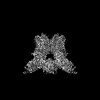
- PDB-8t1d: Open-state cryo-EM structure of full-length human TRPV4 in comple... -
+ Open data

- Basic information
| Entry | Database: PDB / ID: 8t1d | |||||||||||||||||||||
|---|---|---|---|---|---|---|---|---|---|---|---|---|---|---|---|---|---|---|---|---|---|---|
| Title | Open-state cryo-EM structure of full-length human TRPV4 in complex with agonist 4a-PDD | |||||||||||||||||||||
 Components Components | Transient receptor potential cation channel subfamily V member 4/Enhanced green fluorescent protein chimera | |||||||||||||||||||||
 Keywords Keywords | MEMBRANE PROTEIN / transient receptor potential V family member 4 / TRP / channel / TRPV4 / TRP channels / agonist / 4a-PDD / open state / 4alpha-Phorbol 12 / 13-didecanoate | |||||||||||||||||||||
| Function / homology |  Function and homology information Function and homology informationstretch-activated, monoatomic cation-selective, calcium channel activity / blood vessel endothelial cell delamination / osmosensor activity / calcium ion import into cytosol / positive regulation of microtubule depolymerization / positive regulation of macrophage inflammatory protein 1 alpha production / positive regulation of striated muscle contraction / cartilage development involved in endochondral bone morphogenesis / positive regulation of chemokine (C-X-C motif) ligand 1 production / positive regulation of chemokine (C-C motif) ligand 5 production ...stretch-activated, monoatomic cation-selective, calcium channel activity / blood vessel endothelial cell delamination / osmosensor activity / calcium ion import into cytosol / positive regulation of microtubule depolymerization / positive regulation of macrophage inflammatory protein 1 alpha production / positive regulation of striated muscle contraction / cartilage development involved in endochondral bone morphogenesis / positive regulation of chemokine (C-X-C motif) ligand 1 production / positive regulation of chemokine (C-C motif) ligand 5 production / cellular hypotonic salinity response / cellular hypotonic response / cortical microtubule organization / multicellular organismal-level water homeostasis / calcium ion import / positive regulation of vascular permeability / negative regulation of brown fat cell differentiation / positive regulation of monocyte chemotactic protein-1 production / cell-cell junction assembly / cell volume homeostasis / osmosensory signaling pathway / cellular response to osmotic stress / regulation of aerobic respiration / cortical actin cytoskeleton / diet induced thermogenesis / positive regulation of macrophage chemotaxis / TRP channels / microtubule polymerization / calcium ion import across plasma membrane / response to mechanical stimulus / alpha-tubulin binding / beta-tubulin binding / cytoplasmic microtubule / monoatomic cation channel activity / SH2 domain binding / protein kinase C binding / bioluminescence / actin filament organization / filopodium / adherens junction / generation of precursor metabolites and energy / response to insulin / positive regulation of JNK cascade / positive regulation of interleukin-6 production / intracellular calcium ion homeostasis / ruffle membrane / calcium channel activity / calcium ion transmembrane transport / calcium ion transport / actin filament binding / positive regulation of inflammatory response / glucose homeostasis / negative regulation of neuron projection development / lamellipodium / cellular response to heat / positive regulation of cytosolic calcium ion concentration / growth cone / actin cytoskeleton organization / actin binding / High laminar flow shear stress activates signaling by PIEZO1 and PECAM1:CDH5:KDR in endothelial cells / microtubule binding / response to hypoxia / positive regulation of ERK1 and ERK2 cascade / calmodulin binding / cilium / apical plasma membrane / focal adhesion / lipid binding / protein kinase binding / negative regulation of transcription by RNA polymerase II / cell surface / endoplasmic reticulum / ATP binding / membrane / metal ion binding / identical protein binding / plasma membrane Similarity search - Function | |||||||||||||||||||||
| Biological species |  Homo sapiens (human) Homo sapiens (human)  Human betaherpesvirus 5 Human betaherpesvirus 5 | |||||||||||||||||||||
| Method | ELECTRON MICROSCOPY / single particle reconstruction / cryo EM / Resolution: 3.35 Å | |||||||||||||||||||||
 Authors Authors | Talyzina, I.A. / Nadezhdin, K.D. / Neuberger, A. / Sobolevsky, A.I. | |||||||||||||||||||||
| Funding support |  United States, United States,  Germany, 6items Germany, 6items
| |||||||||||||||||||||
 Citation Citation | Journal: Nat Commun / Year: 2023 Title: Structure of human TRPV4 in complex with GTPase RhoA. Authors: Kirill D Nadezhdin / Irina A Talyzina / Aravind Parthasarathy / Arthur Neuberger / David X Zhang / Alexander I Sobolevsky / Abstract: Transient receptor potential (TRP) channel TRPV4 is a polymodal cellular sensor that responds to moderate heat, cell swelling, shear stress, and small-molecule ligands. It is involved in ...Transient receptor potential (TRP) channel TRPV4 is a polymodal cellular sensor that responds to moderate heat, cell swelling, shear stress, and small-molecule ligands. It is involved in thermogenesis, regulation of vascular tone, bone homeostasis, renal and pulmonary functions. TRPV4 is implicated in neuromuscular and skeletal disorders, pulmonary edema, and cancers, and represents an important drug target. The cytoskeletal remodeling GTPase RhoA has been shown to suppress TRPV4 activity. Here, we present a structure of the human TRPV4-RhoA complex that shows RhoA interaction with the membrane-facing surface of the TRPV4 ankyrin repeat domains. The contact interface reveals residues that are mutated in neuropathies, providing an insight into the disease pathogenesis. We also identify the binding sites of the TRPV4 agonist 4α-PDD and the inhibitor HC-067047 at the base of the S1-S4 bundle, and show that agonist binding leads to pore opening, while channel inhibition involves a π-to-α transition in the pore-forming helix S6. Our structures elucidate the interaction interface between hTRPV4 and RhoA, as well as residues at this interface that are involved in TRPV4 disease-causing mutations. They shed light on TRPV4 activation and inhibition and provide a template for the design of future therapeutics for treatment of TRPV4-related diseases. | |||||||||||||||||||||
| History |
|
- Structure visualization
| Structure viewer | Molecule: MolmilJmol/JSmol |
|---|
- Downloads & links
-Download
| PDBx/mmCIF format | 8t1d.cif.gz | 481.2 KB | Display | PDBx/mmCIF format |
|---|---|---|---|---|
| PDB format | pdb8t1d.ent.gz | 373.8 KB | Display | PDB format |
| PDBx/mmJSON format | 8t1d.json.gz | Tree view | PDBx/mmJSON format | |
| Others |  Other downloads Other downloads |
-Validation report
| Arichive directory | https://data.pdbj.org/pub/pdb/validation_reports/t1/8t1dftp://data.pdbj.org/pub/pdb/validation_reports/t1/8t1d | HTTPS FTP |
|---|
-Related structure data
| Related structure data | 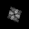 40960MC 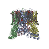 8t1bC  8t1cC 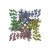 8t1eC  8t1fC C: citing same article ( M: map data used to model this data |
|---|---|
| Similar structure data |


-Links
 PDBj
PDBj


- Assembly
| Deposited unit | 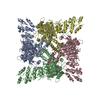
|
|---|---|
| 1 |
|
-Components
| #1: Protein | Mass: 127717.938 Da / Num. of mol.: 4 Source method: isolated from a genetically manipulated source Source: (gene. exp.) Homo sapiens (human), (gene. exp.) Human betaherpesvirus 5Gene: TRPV4, VRL2, VROAC, egfp / Cell line (production host): HEK293S / Production host: Homo sapiens (human) / References: UniProt: Q9HBA0, UniProt: C5MKY7#2: Chemical | ChemComp-XS9 / ( 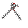  Mass: 672.931 Da / Num. of mol.: 4 / Source method: obtained synthetically / Formula: C40H64O8 / Feature type: SUBJECT OF INVESTIGATION Mass: 672.931 Da / Num. of mol.: 4 / Source method: obtained synthetically / Formula: C40H64O8 / Feature type: SUBJECT OF INVESTIGATIONHas ligand of interest | Y | Has protein modification | Y | |
|---|
-Experimental details
-Experiment
| Experiment | Method: ELECTRON MICROSCOPY |
|---|---|
| EM experiment | Aggregation state: PARTICLE / 3D reconstruction method: single particle reconstruction |
- Sample preparation
| Component | Name: full-length human TRPV4 in complex with agonist 4a-PDD Type: COMPLEX / Entity ID: #1 / Source: RECOMBINANT | ||||||||||||||||||||||||||||||
|---|---|---|---|---|---|---|---|---|---|---|---|---|---|---|---|---|---|---|---|---|---|---|---|---|---|---|---|---|---|---|---|
| Molecular weight | Value: 0.51 MDa / Experimental value: NO | ||||||||||||||||||||||||||||||
| Source (natural) | Organism: Homo sapiens (human) | ||||||||||||||||||||||||||||||
| Source (recombinant) | Organism: Homo sapiens (human) / Cell: Human embryonic kidney 293 / Plasmid: pEG BacMam | ||||||||||||||||||||||||||||||
| Buffer solution | pH: 8 | ||||||||||||||||||||||||||||||
| Buffer component |
| ||||||||||||||||||||||||||||||
| Specimen | Conc.: 3.2 mg/ml / Embedding applied: NO / Shadowing applied: NO / Staining applied: NO / Vitrification applied: YES / Details: human TRPV4 | ||||||||||||||||||||||||||||||
| Specimen support | Grid material: GOLD / Grid mesh size: 300 divisions/in. / Grid type: UltrAuFoil R1.2/1.3 | ||||||||||||||||||||||||||||||
| Vitrification | Instrument: FEI VITROBOT MARK IV / Cryogen name: ETHANE / Humidity: 100 % / Chamber temperature: 295 K |
- Electron microscopy imaging
| Experimental equipment |  Model: Titan Krios / Image courtesy: FEI Company |
|---|---|
| Microscopy | Model: TFS KRIOS |
| Electron gun | Electron source:  FIELD EMISSION GUN / Accelerating voltage: 300 kV / Illumination mode: FLOOD BEAM FIELD EMISSION GUN / Accelerating voltage: 300 kV / Illumination mode: FLOOD BEAM |
| Electron lens | Mode: BRIGHT FIELD / Nominal defocus max: 1500 nm / Nominal defocus min: 750 nm / Cs: 2.7 mm |
| Image recording | Average exposure time: 2.4 sec. / Electron dose: 60 e/Å2 / Film or detector model: GATAN K3 (6k x 4k) / Num. of grids imaged: 1 / Num. of real images: 15624 |
| Image scans | Width: 11520 / Height: 8184 |
- Processing
| EM software | Name: PHENIX / Version: 1.11.1_2575: / Category: model refinement | ||||||||||||||||||||||||
|---|---|---|---|---|---|---|---|---|---|---|---|---|---|---|---|---|---|---|---|---|---|---|---|---|---|
| CTF correction | Type: NONE | ||||||||||||||||||||||||
| Particle selection | Num. of particles selected: 3935964 | ||||||||||||||||||||||||
| 3D reconstruction | Resolution: 3.35 Å / Resolution method: FSC 0.143 CUT-OFF / Num. of particles: 80823 / Symmetry type: POINT | ||||||||||||||||||||||||
| Atomic model building | Space: REAL | ||||||||||||||||||||||||
| Refine LS restraints |
|
