 Movie
Movie Controller

[English] 日本語
 Yorodumi
- PDB-8szy: Crystal Structure of Heterotrimeric Anti-TIGIT Fabs in complex wi... -
+ Open data

- Basic information
| Entry | Database: PDB / ID: 8szy | ||||||
|---|---|---|---|---|---|---|---|
| Title | Crystal Structure of Heterotrimeric Anti-TIGIT Fabs in complex with human TIGIT | ||||||
 Components Components |
| ||||||
 Keywords Keywords | IMMUNE SYSTEM / PROTEIN BINDING / Antibody / TIGIT / PVR-Family | ||||||
| Function / homology |  Function and homology information Function and homology informationnegative regulation of T cell activation / negative regulation of interleukin-12 production / negative regulation of natural killer cell mediated cytotoxicity / positive regulation of interleukin-10 production / signaling receptor activity / signaling receptor binding / cell surface / identical protein binding / plasma membrane Similarity search - Function | ||||||
| Biological species |  Homo sapiens (human) Homo sapiens (human) | ||||||
| Method |  X-RAY DIFFRACTION / SYNCHROTRON / MOLECULAR REPLACEMENT / Resolution: 2.31 Å X-RAY DIFFRACTION / SYNCHROTRON / MOLECULAR REPLACEMENT / Resolution: 2.31 Å | ||||||
 Authors Authors | Diong, S.J. / Lee, P.S. | ||||||
| Funding support |  United States, 1items United States, 1items
| ||||||
 Citation Citation | Journal: Mabs / Year: 2023 Title: Biophysical characterization of PVR family interactions and therapeutic antibody recognition to TIGIT. Authors: Diong, S.J. / Jashnani, A. / Drake, A.W. / Bee, C. / Findeisen, F. / Dollinger, G. / Wang, F. / Rajpal, A. / Strop, P. / Lee, P.S. | ||||||
| History |
|
- Structure visualization
| Structure viewer | Molecule: MolmilJmol/JSmol |
|---|
- Downloads & links
-Download
| PDBx/mmCIF format | 8szy.cif.gz | 1.1 MB | Display | PDBx/mmCIF format |
|---|---|---|---|---|
| PDB format | pdb8szy.ent.gz | 866.4 KB | Display | PDB format |
| PDBx/mmJSON format | 8szy.json.gz | Tree view | PDBx/mmJSON format | |
| Others |  Other downloads Other downloads |
-Validation report
| Summary document | 8szy_validation.pdf.gz | 528.6 KB | Display | wwPDB validaton report |
|---|---|---|---|---|
| Full document | 8szy_full_validation.pdf.gz | 544.7 KB | Display | |
| Data in XML | 8szy_validation.xml.gz | 65.2 KB | Display | |
| Data in CIF | 8szy_validation.cif.gz | 89.7 KB | Display | |
| Arichive directory | https://data.pdbj.org/pub/pdb/validation_reports/sz/8szyftp://data.pdbj.org/pub/pdb/validation_reports/sz/8szy | HTTPS FTP |
-Related structure data
| Similar structure data |
|---|
-Links
 PDBj
PDBj

- Assembly
| Deposited unit | 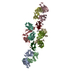
| ||||||||||
|---|---|---|---|---|---|---|---|---|---|---|---|
| 1 | 
| ||||||||||
| 2 | 
| ||||||||||
| Unit cell |
|
-Components
-Antibody , 4 types, 8 molecules ACBDHILM
| #1: Antibody | Mass: 24309.189 Da / Num. of mol.: 2 Source method: isolated from a genetically manipulated source Source: (gene. exp.) Homo sapiens (human) / Production host: Homo sapiens (human)#2: Antibody | Mass: 24072.754 Da / Num. of mol.: 2 Source method: isolated from a genetically manipulated source Source: (gene. exp.) Homo sapiens (human) / Production host: Homo sapiens (human)#3: Antibody | Mass: 25870.719 Da / Num. of mol.: 2 Source method: isolated from a genetically manipulated source Source: (gene. exp.) Homo sapiens (human) / Cell line (production host): Expi293F / Production host: Homo sapiens (human)#4: Antibody | Mass: 23621.234 Da / Num. of mol.: 2 Source method: isolated from a genetically manipulated source Source: (gene. exp.) Homo sapiens (human) / Production host: Homo sapiens (human) |
|---|
-Protein / Sugars / Non-polymers , 3 types, 116 molecules TU

| #5: Protein | Mass: 12483.785 Da / Num. of mol.: 2 / Mutation: C69S Source method: isolated from a genetically manipulated source Source: (gene. exp.) Homo sapiens (human) / Gene: TIGIT, VSIG9, VSTM3 / Production host: Homo sapiens (human) / References: UniProt: Q495A1#6: Sugar |  Type: D-saccharide, beta linking / Mass: 221.208 Da / Num. of mol.: 2 / Source method: isolated from a natural source / Formula: C8H15NO6 Type: D-saccharide, beta linking / Mass: 221.208 Da / Num. of mol.: 2 / Source method: isolated from a natural source / Formula: C8H15NO6#7: Water | ChemComp-HOH / | Mass: 18.015 Da / Num. of mol.: 112 / Source method: isolated from a natural source / Formula: H2O |
|---|
-Details
| Has ligand of interest | N |
|---|---|
| Has protein modification | Y |
-Experimental details
-Experiment
| Experiment | Method: X-RAY DIFFRACTION / Number of used crystals: 1 |
|---|
- Sample preparation
| Crystal | Density Matthews: 2.45 Å3/Da / Density % sol: 49.75 % |
|---|---|
| Crystal grow | Temperature: 300 K / Method: vapor diffusion, sitting drop Details: 0.2 M sodium phosphate monobasic monohydrate, 20% w/v Polyethylene glycol 1,000 |
-Data collection
| Diffraction | Mean temperature: 100 K / Serial crystal experiment: N |
|---|---|
| Diffraction source | Source: SYNCHROTRON / Site: APS / Beamline: 17-ID / Wavelength: 1 Å |
| Detector | Type: DECTRIS PILATUS 6M / Detector: PIXEL / Date: Feb 19, 2020 |
| Radiation | Protocol: SINGLE WAVELENGTH / Monochromatic (M) / Laue (L): M / Scattering type: x-ray |
| Radiation wavelength | Wavelength: 1 Å / Relative weight: 1 |
| Reflection | Resolution: 2.306→108.856 Å / Num. obs: 65410 / % possible obs: 70.2 % / Redundancy: 3.3 % / Biso Wilson estimate: 35.04 Å2 / CC1/2: 0.985 / Rmerge(I) obs: 0.14 / Rpim(I) all: 0.091 / Rrim(I) all: 0.168 / Net I/σ(I): 7.5 |
| Reflection shell | Resolution: 2.306→2.479 Å / Rmerge(I) obs: 0.694 / Mean I/σ(I) obs: 1.5 / Num. unique obs: 3271 / CC1/2: 0.6 / Rpim(I) all: 0.491 / Rrim(I) all: 0.854 |
- Processing
| Software |
| ||||||||||||||||||||||||||||||||||||||||||||||||||||||||||||||||||||||||||||||||||||||||||||||||||||||||||||||||||||||||||||||||||||||||||||||||||||||||||||||||||||||||
|---|---|---|---|---|---|---|---|---|---|---|---|---|---|---|---|---|---|---|---|---|---|---|---|---|---|---|---|---|---|---|---|---|---|---|---|---|---|---|---|---|---|---|---|---|---|---|---|---|---|---|---|---|---|---|---|---|---|---|---|---|---|---|---|---|---|---|---|---|---|---|---|---|---|---|---|---|---|---|---|---|---|---|---|---|---|---|---|---|---|---|---|---|---|---|---|---|---|---|---|---|---|---|---|---|---|---|---|---|---|---|---|---|---|---|---|---|---|---|---|---|---|---|---|---|---|---|---|---|---|---|---|---|---|---|---|---|---|---|---|---|---|---|---|---|---|---|---|---|---|---|---|---|---|---|---|---|---|---|---|---|---|---|---|---|---|---|---|---|---|
| Refinement | Method to determine structure: MOLECULAR REPLACEMENT / Resolution: 2.31→45.6 Å / SU ML: 0.3176 / Cross valid method: FREE R-VALUE / σ(F): 1.36 / Phase error: 33.0909 Stereochemistry target values: GeoStd + Monomer Library + CDL v1.2
| ||||||||||||||||||||||||||||||||||||||||||||||||||||||||||||||||||||||||||||||||||||||||||||||||||||||||||||||||||||||||||||||||||||||||||||||||||||||||||||||||||||||||
| Solvent computation | Shrinkage radii: 0.9 Å / VDW probe radii: 1.11 Å / Solvent model: FLAT BULK SOLVENT MODEL | ||||||||||||||||||||||||||||||||||||||||||||||||||||||||||||||||||||||||||||||||||||||||||||||||||||||||||||||||||||||||||||||||||||||||||||||||||||||||||||||||||||||||
| Displacement parameters | Biso mean: 34.66 Å2 | ||||||||||||||||||||||||||||||||||||||||||||||||||||||||||||||||||||||||||||||||||||||||||||||||||||||||||||||||||||||||||||||||||||||||||||||||||||||||||||||||||||||||
| Refinement step | Cycle: LAST / Resolution: 2.31→45.6 Å
| ||||||||||||||||||||||||||||||||||||||||||||||||||||||||||||||||||||||||||||||||||||||||||||||||||||||||||||||||||||||||||||||||||||||||||||||||||||||||||||||||||||||||
| Refine LS restraints |
| ||||||||||||||||||||||||||||||||||||||||||||||||||||||||||||||||||||||||||||||||||||||||||||||||||||||||||||||||||||||||||||||||||||||||||||||||||||||||||||||||||||||||
| LS refinement shell |
| ||||||||||||||||||||||||||||||||||||||||||||||||||||||||||||||||||||||||||||||||||||||||||||||||||||||||||||||||||||||||||||||||||||||||||||||||||||||||||||||||||||||||
| Refinement TLS params. | Method: refined / Origin x: 11.5172330803 Å / Origin y: 39.7083482266 Å / Origin z: -26.4063142197 Å
| ||||||||||||||||||||||||||||||||||||||||||||||||||||||||||||||||||||||||||||||||||||||||||||||||||||||||||||||||||||||||||||||||||||||||||||||||||||||||||||||||||||||||
| Refinement TLS group | Selection details: all |
