 ムービー
ムービー コントローラー


+ データを開く

- 基本情報
基本情報
| 登録情報 | データベース: PDB / ID: 8syf | ||||||||||||||||||||||||||||||
|---|---|---|---|---|---|---|---|---|---|---|---|---|---|---|---|---|---|---|---|---|---|---|---|---|---|---|---|---|---|---|---|
| タイトル | Homology model of Acto-HMM complex in ADP-state. Chicken smooth muscle HMM and chicken pectoralis actin | ||||||||||||||||||||||||||||||
 要素 要素 |
| ||||||||||||||||||||||||||||||
 キーワード キーワード | MOTOR PROTEIN / Actin / Myosin / Smooth muscle / Cryo-EM / Cryo-ET / Heavy meromyosin | ||||||||||||||||||||||||||||||
| 機能・相同性 |  機能・相同性情報 機能・相同性情報RHO GTPases activate PAKs / myosin II filament / Smooth Muscle Contraction / myofibril assembly / myosin II binding / Striated Muscle Contraction / muscle myosin complex / actomyosin / actomyosin structure organization / myosin complex ...RHO GTPases activate PAKs / myosin II filament / Smooth Muscle Contraction / myofibril assembly / myosin II binding / Striated Muscle Contraction / muscle myosin complex / actomyosin / actomyosin structure organization / myosin complex / myosin II complex / structural constituent of muscle / microfilament motor activity / myosin heavy chain binding / myofibril / cytoskeletal motor activity / striated muscle thin filament / skeletal muscle thin filament assembly / skeletal muscle fiber development / stress fiber / actin filament / ADP binding / 加水分解酵素; 酸無水物に作用; 酸無水物に作用・細胞または細胞小器官の運動に関与 / actin filament binding / actin cytoskeleton / hydrolase activity / calcium ion binding / magnesium ion binding / ATP binding / cytosol / cytoplasm 類似検索 - 分子機能 | ||||||||||||||||||||||||||||||
| 生物種 |  | ||||||||||||||||||||||||||||||
| 手法 | 電子顕微鏡法 / 単粒子再構成法 / クライオ電子顕微鏡法 / 解像度: 19 Å | ||||||||||||||||||||||||||||||
 データ登録者 データ登録者 | Hojjatian, A. / Taylor, D.W. / Daneshparvar, N. / Trybus, K.M. / Taylor, K.A. | ||||||||||||||||||||||||||||||
| 資金援助 |  米国, 9件 米国, 9件
| ||||||||||||||||||||||||||||||
 引用 引用 | ジャーナル: J Struct Biol / 年: 2023 タイトル: Double-headed binding of myosin II to F-actin shows the effect of strain on head structure. 著者: Alimohammad Hojjatian / Dianne W Taylor / Nadia Daneshparvar / Patricia M Fagnant / Kathleen M Trybus / Kenneth A Taylor / 要旨: Force production in muscle is achieved through the interaction of myosin and actin. Strong binding states in active muscle are associated with Mg·ADP bound to the active site; release of Mg·ADP ...Force production in muscle is achieved through the interaction of myosin and actin. Strong binding states in active muscle are associated with Mg·ADP bound to the active site; release of Mg·ADP allows rebinding of ATP and dissociation from actin. Thus, Mg·ADP binding is positioned for adaptation as a force sensor. Mechanical loads on the lever arm can affect the ability of myosin to release Mg·ADP but exactly how this is done is poorly defined. Here we use F-actin decorated with double-headed smooth muscle myosin fragments in the presence of Mg·ADP to visualize the effect of internally supplied tension on the paired lever arms using cryoEM. The interaction of the paired heads with two adjacent actin subunits is predicted to place one lever arm under positive and the other under negative strain. The converter domain is believed to be the most flexible domain within myosin head. Our results, instead, point to the segment of heavy chain between the essential and regulatory light chains as the location of the largest structural change. Moreover, our results suggest no large changes in the myosin coiled coil tail as the locus of strain relief when both heads bind F-actin. The method would be adaptable to double-headed members of the myosin family. We anticipate that the study of actin-myosin interaction using double-headed fragments enables visualization of domains that are typically noisy in decoration with single-headed fragments. | ||||||||||||||||||||||||||||||
| 履歴 |
|
- 構造の表示
構造の表示
| 構造ビューア | 分子: MolmilJmol/JSmol |
|---|
- ダウンロードとリンク
-ダウンロード
| PDBx/mmCIF形式 | 8syf.cif.gz | 515.6 KB | 表示 | PDBx/mmCIF形式 |
|---|---|---|---|---|
| PDB形式 | pdb8syf.ent.gz | 415.7 KB | 表示 | PDB形式 |
| PDBx/mmJSON形式 | 8syf.json.gz | ツリー表示 | PDBx/mmJSON形式 | |
| その他 |  その他のダウンロード その他のダウンロード |
-検証レポート
| 文書・要旨 | 8syf_validation.pdf.gz | 1.1 MB | 表示 | wwPDB検証レポート |
|---|---|---|---|---|
| 文書・詳細版 | 8syf_full_validation.pdf.gz | 1.5 MB | 表示 | |
| XML形式データ | 8syf_validation.xml.gz | 139.3 KB | 表示 | |
| CIF形式データ | 8syf_validation.cif.gz | 207.4 KB | 表示 | |
| アーカイブディレクトリ | https://data.pdbj.org/pub/pdb/validation_reports/sy/8syfftp://data.pdbj.org/pub/pdb/validation_reports/sy/8syf | HTTPS FTP |
-関連構造データ
| 関連構造データ | 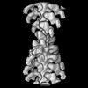 29646MC M: このデータのモデリングに利用したマップデータ C: 同じ文献を引用 ( |
|---|---|
| 類似構造データ | |
| 実験データセット #1 | データ参照: 10.6019/EMPIAR-11555 / データの種類: EMPIAR |
-リンク
 PDBj
PDBj


- 集合体
集合体
| 登録構造単位 | 
|
|---|---|
| 1 |
|
-要素
| #1: タンパク質 | 分子量: 96768.078 Da / 分子数: 2 / 由来タイプ: 組換発現 / 由来: (組換発現)  unidentified baculovirus (ウイルス) / 参照: UniProt: A0A8V0ZE13 unidentified baculovirus (ウイルス) / 参照: UniProt: A0A8V0ZE13#2: タンパク質 | 分子量: 41862.613 Da / 分子数: 2 / 由来タイプ: 天然 / 由来: (天然) 参照: UniProt: P68139, 加水分解酵素; 酸無水物に作用; 酸無水物に作用・細胞または細胞小器官の運動に関与 #3: タンパク質 | 分子量: 16639.715 Da / 分子数: 2 / 由来タイプ: 組換発現 / 由来: (組換発現) unidentified baculovirus (ウイルス) / 参照: UniProt: P02607#4: タンパク質 | 分子量: 16583.412 Da / 分子数: 2 / 由来タイプ: 組換発現 / 由来: (組換発現) unidentified baculovirus (ウイルス) / 参照: UniProt: P02612Has protein modification | N | |
|---|
-実験情報
-実験
| 実験 | 手法: 電子顕微鏡法 |
|---|---|
| EM実験 | 試料の集合状態: FILAMENT / 3次元再構成法: 単粒子再構成法 |
- 試料調製
試料調製
| 構成要素 | 名称: Filamentous actin decorated with chicken smooth muscle heavy meromyosin タイプ: COMPLEX / Entity ID: all / 由来: RECOMBINANT |
|---|---|
| 分子量 | 実験値: NO |
| 由来(天然) | 生物種: |
| 由来(組換発現) | 生物種: unidentified baculovirus (ウイルス) |
| 緩衝液 | pH: 7.6 |
| 試料 | 包埋: NO / シャドウイング: NO / 染色: NO / 凍結: YES |
| 急速凍結 | 凍結剤: ETHANE-PROPANE |
- 電子顕微鏡撮影
| 実験機器 |  モデル: Titan Krios / 画像提供: FEI Company |
|---|---|
| 顕微鏡 | モデル: FEI TITAN KRIOS |
| 電子銃 | 電子線源:  FIELD EMISSION GUN / 加速電圧: 300 kV / 照射モード: OTHER FIELD EMISSION GUN / 加速電圧: 300 kV / 照射モード: OTHER |
| 電子レンズ | モード: BRIGHT FIELD / 最大 デフォーカス(公称値): 2000 nm / 最小 デフォーカス(公称値): 200 nm |
| 撮影 | 電子線照射量: 29.6 e/Å2 / 検出モード: INTEGRATING フィルム・検出器のモデル: DIRECT ELECTRON DE-64 (8k x 8k) |
- 解析
解析
| EMソフトウェア | 名称: PHENIX / カテゴリ: モデル精密化 | ||||||||||||||||||||||||
|---|---|---|---|---|---|---|---|---|---|---|---|---|---|---|---|---|---|---|---|---|---|---|---|---|---|
| CTF補正 | タイプ: NONE | ||||||||||||||||||||||||
| 3次元再構成 | 解像度: 19 Å / 解像度の算出法: FSC 0.143 CUT-OFF / 粒子像の数: 17394 / 対称性のタイプ: POINT | ||||||||||||||||||||||||
| 拘束条件 |
|
