+ Open data
- Basic information
| Entry | Database: PDB / ID: 8sp3 | ||||||
|---|---|---|---|---|---|---|---|
| Title | Asymmetric dimer of MapSPARTA bound with gRNA/tDNA hybrid | ||||||
 Components Components |
| ||||||
 Keywords Keywords | IMMUNE SYSTEM / Short prokaryotic argonaute / Oligomerization / TIR / NADase activity / Bacterial immune system / MapSPARTA | ||||||
| Function / homology |  Function and homology information Function and homology information | ||||||
| Biological species |  Maribacter polysiphoniae (bacteria) Maribacter polysiphoniae (bacteria) | ||||||
| Method | ELECTRON MICROSCOPY / single particle reconstruction / cryo EM / Resolution: 3.52 Å | ||||||
 Authors Authors | Shen, Z.F. / Yang, X.Y. / Fu, T.M. | ||||||
| Funding support |  United States, 1items United States, 1items
| ||||||
 Citation Citation |  Journal: Nature / Year: 2023 Journal: Nature / Year: 2023Title: Oligomerization-mediated activation of a short prokaryotic Argonaute. Authors: Zhangfei Shen / Xiao-Yuan Yang / Shiyu Xia / Wei Huang / Derek J Taylor / Kotaro Nakanishi / Tian-Min Fu /  Abstract: Although eukaryotic and long prokaryotic Argonaute proteins (pAgos) cleave nucleic acids, some short pAgos lack nuclease activity and hydrolyse NAD(P) to induce bacterial cell death. Here we present ...Although eukaryotic and long prokaryotic Argonaute proteins (pAgos) cleave nucleic acids, some short pAgos lack nuclease activity and hydrolyse NAD(P) to induce bacterial cell death. Here we present a hierarchical activation pathway for SPARTA, a short pAgo consisting of an Argonaute (Ago) protein and TIR-APAZ, an associated protein. SPARTA progresses through distinct oligomeric forms, including a monomeric apo state, a monomeric RNA-DNA-bound state, two dimeric RNA-DNA-bound states and a tetrameric RNA-DNA-bound active state. These snapshots together identify oligomerization as a mechanistic principle of SPARTA activation. The RNA-DNA-binding channel of apo inactive SPARTA is occupied by an auto-inhibitory motif in TIR-APAZ. After the binding of RNA-DNA, SPARTA transitions from a monomer to a symmetric dimer and then an asymmetric dimer, in which two TIR domains interact through charge and shape complementarity. Next, two dimers assemble into a tetramer with a central TIR cluster responsible for hydrolysing NAD(P). In addition, we observe unique features of interactions between SPARTA and RNA-DNA, including competition between the DNA 3' end and the auto-inhibitory motif, interactions between the RNA G2 nucleotide and Ago, and splaying of the RNA-DNA duplex by two loops exclusive to short pAgos. Together, our findings provide a mechanistic basis for the activation of short pAgos, a large section of the Ago superfamily. | ||||||
| History |
|
- Structure visualization
| Structure viewer | Molecule:  Molmil Molmil Jmol/JSmol Jmol/JSmol |
|---|
- Downloads & links
- Download
| PDBx/mmCIF format |  8sp3.cif.gz 8sp3.cif.gz | 372 KB | Display |  PDBx/mmCIF format PDBx/mmCIF format |
|---|---|---|---|---|
| PDB format |  pdb8sp3.ent.gz pdb8sp3.ent.gz | 295.1 KB | Display |  PDB format PDB format |
| PDBx/mmJSON format |  8sp3.json.gz 8sp3.json.gz | Tree view |  PDBx/mmJSON format PDBx/mmJSON format | |
| Others |  Other downloads Other downloads |
-Validation report
| Arichive directory |  https://data.pdbj.org/pub/pdb/validation_reports/sp/8sp3 https://data.pdbj.org/pub/pdb/validation_reports/sp/8sp3 ftp://data.pdbj.org/pub/pdb/validation_reports/sp/8sp3 ftp://data.pdbj.org/pub/pdb/validation_reports/sp/8sp3 | HTTPS FTP |
|---|
-Related structure data
| Related structure data |  40673MC 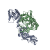 8fexC 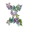 8ffiC 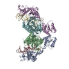 8sp0C 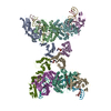 8spoC 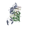 8squC M: map data used to model this data C: citing same article ( |
|---|---|
| Similar structure data | Similarity search - Function & homology  F&H Search F&H Search |
- Links
Links
- Assembly
| Deposited unit | 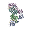
|
|---|---|
| 1 |
|
- Components
| #1: Protein | Mass: 53139.398 Da / Num. of mol.: 2 Source method: isolated from a genetically manipulated source Source: (gene. exp.)  Maribacter polysiphoniae (bacteria) / Gene: LX92_01810 / Production host: Maribacter polysiphoniae (bacteria) / Gene: LX92_01810 / Production host:  #2: Protein | Mass: 58091.410 Da / Num. of mol.: 2 Source method: isolated from a genetically manipulated source Source: (gene. exp.)  Maribacter polysiphoniae (bacteria) / Gene: LX92_01809 / Production host: Maribacter polysiphoniae (bacteria) / Gene: LX92_01809 / Production host:  #3: RNA chain | Mass: 6651.949 Da / Num. of mol.: 2 / Source method: obtained synthetically / Source: (synth.)  Maribacter polysiphoniae (bacteria) Maribacter polysiphoniae (bacteria)#4: DNA chain | Mass: 7675.000 Da / Num. of mol.: 2 / Source method: obtained synthetically / Source: (synth.)  Maribacter polysiphoniae (bacteria) Maribacter polysiphoniae (bacteria)#5: Chemical | Has ligand of interest | Y | |
|---|
-Experimental details
-Experiment
| Experiment | Method: ELECTRON MICROSCOPY |
|---|---|
| EM experiment | Aggregation state: PARTICLE / 3D reconstruction method: single particle reconstruction |
- Sample preparation
| Component | Name: Asymmetric dimer of MapSPARTA bound with gRNA/tDNA hybrid Type: COMPLEX / Entity ID: #1-#4 / Source: RECOMBINANT |
|---|---|
| Source (natural) | Organism:  Maribacter polysiphoniae (bacteria) Maribacter polysiphoniae (bacteria) |
| Source (recombinant) | Organism:  |
| Buffer solution | pH: 8 |
| Specimen | Embedding applied: NO / Shadowing applied: NO / Staining applied: NO / Vitrification applied: YES |
| Vitrification | Cryogen name: ETHANE |
- Electron microscopy imaging
| Experimental equipment |  Model: Titan Krios / Image courtesy: FEI Company |
|---|---|
| Microscopy | Model: FEI TITAN KRIOS |
| Electron gun | Electron source:  FIELD EMISSION GUN / Accelerating voltage: 300 kV / Illumination mode: FLOOD BEAM FIELD EMISSION GUN / Accelerating voltage: 300 kV / Illumination mode: FLOOD BEAM |
| Electron lens | Mode: BRIGHT FIELD / Nominal defocus max: 2100 nm / Nominal defocus min: 500 nm |
| Image recording | Electron dose: 50 e/Å2 / Film or detector model: FEI FALCON IV (4k x 4k) |
- Processing
| CTF correction | Type: NONE |
|---|---|
| 3D reconstruction | Resolution: 3.52 Å / Resolution method: FSC 0.143 CUT-OFF / Num. of particles: 32366 / Symmetry type: POINT |
 Movie
Movie Controller


 PDBj
PDBj


