 Movie
Movie Controller


+ Open data

- Basic information
| Entry | Database: PDB / ID: 8smc | ||||||
|---|---|---|---|---|---|---|---|
| Title | Cryo-EM structure of LRRK2 bound with type-I inhibitor DNL201 | ||||||
 Components Components | non-specific serine/threonine protein kinase | ||||||
 Keywords Keywords | HYDROLASE / Parkinson's Disease / kinase / activation | ||||||
| Function / homology |  Function and homology information Function and homology informationregulation of multicellular organismal process / intracellular organelle lumen / Wnt signalosome assembly / GTP-dependent protein kinase activity / protein localization to endoplasmic reticulum exit site / animal organ development / regulation of intracellular signal transduction / regulation of dopamine receptor signaling pathway / regulation of vesicle-mediated transport / regulation of dendritic spine morphogenesis ...regulation of multicellular organismal process / intracellular organelle lumen / Wnt signalosome assembly / GTP-dependent protein kinase activity / protein localization to endoplasmic reticulum exit site / animal organ development / regulation of intracellular signal transduction / regulation of dopamine receptor signaling pathway / regulation of vesicle-mediated transport / regulation of dendritic spine morphogenesis / endoplasmic reticulum organization / Wnt signalosome / regulation of canonical Wnt signaling pathway / Golgi organization / protein kinase A binding / exploration behavior / positive regulation of protein metabolic process / regulation of proteolysis / endoplasmic reticulum exit site / anatomical structure morphogenesis / negative regulation of signal transduction / phagocytic vesicle / vesicle-mediated transport / positive regulation of autophagy / GTPase activator activity / regulation of membrane potential / modulation of chemical synaptic transmission / autophagy / small GTPase binding / neuron differentiation / terminal bouton / protein autophosphorylation / synaptic vesicle membrane / signaling receptor complex adaptor activity / regulation of gene expression / cytoskeleton / protein phosphorylation / perikaryon / mitochondrial outer membrane / non-specific serine/threonine protein kinase / cell surface receptor signaling pathway / lysosome / endosome / intracellular signal transduction / membrane raft / Golgi membrane / hydrolase activity / dendrite / endoplasmic reticulum membrane / GTP binding / ATP binding / identical protein binding / plasma membrane / cytosol Similarity search - Function | ||||||
| Biological species |  Homo sapiens (human) Homo sapiens (human) | ||||||
| Method | ELECTRON MICROSCOPY / single particle reconstruction / cryo EM / Resolution: 4.02 Å | ||||||
 Authors Authors | Sun, J. / Zhu, H. | ||||||
| Funding support |  United States, 1items United States, 1items
| ||||||
 Citation Citation | Journal: Science / Year: 2023 Title: Rab29-dependent asymmetrical activation of leucine-rich repeat kinase 2. Authors: Hanwen Zhu / Francesca Tonelli / Martin Turk / Alan Prescott / Dario R Alessi / Ji Sun /  Abstract: Gain-of-function mutations in , which encodes the leucine-rich repeat kinase 2 (LRRK2), are the most common genetic cause of late-onset Parkinson's disease. LRRK2 is recruited to membrane organelles ...Gain-of-function mutations in , which encodes the leucine-rich repeat kinase 2 (LRRK2), are the most common genetic cause of late-onset Parkinson's disease. LRRK2 is recruited to membrane organelles and activated by Rab29, a Rab guanosine triphosphatase encoded in the locus. We present cryo-electron microscopy structures of Rab29-LRRK2 complexes in three oligomeric states, providing key snapshots during LRRK2 recruitment and activation. Rab29 induces an unexpected tetrameric assembly of LRRK2, formed by two kinase-active central protomers and two kinase-inactive peripheral protomers. The central protomers resemble the active-like state trapped by the type I kinase inhibitor DNL201, a compound that underwent a phase 1 clinical trial. Our work reveals the structural mechanism of LRRK2 spatial regulation and provides insights into LRRK2 inhibitor design for Parkinson's disease treatment. | ||||||
| History |
|
- Structure visualization
| Structure viewer | Molecule: MolmilJmol/JSmol |
|---|
- Downloads & links
-Download
| PDBx/mmCIF format | 8smc.cif.gz | 197.6 KB | Display | PDBx/mmCIF format |
|---|---|---|---|---|
| PDB format | pdb8smc.ent.gz | 144.3 KB | Display | PDB format |
| PDBx/mmJSON format | 8smc.json.gz | Tree view | PDBx/mmJSON format | |
| Others |  Other downloads Other downloads |
-Validation report
| Arichive directory | https://data.pdbj.org/pub/pdb/validation_reports/sm/8smcftp://data.pdbj.org/pub/pdb/validation_reports/sm/8smc | HTTPS FTP |
|---|
-Related structure data
| Related structure data |  40588MC  8fo2C  8fo8C 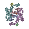 8fo9C M: map data used to model this data C: citing same article ( |
|---|---|
| Similar structure data |
-Links
 PDBj
PDBj


- Assembly
| Deposited unit | 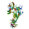
|
|---|---|
| 1 |
|
-Components
| #1: Protein | Mass: 136843.469 Da / Num. of mol.: 1 Source method: isolated from a genetically manipulated source Source: (gene. exp.) Homo sapiens (human) / Gene: LRRK2 / Production host: Homo sapiens (human) / References: UniProt: Q17RV3 |
|---|---|
| #2: Chemical | ChemComp-GDP /   Type: RNA linking / Mass: 443.201 Da / Num. of mol.: 1 / Source method: obtained synthetically / Formula: C10H15N5O11P2 / Feature type: SUBJECT OF INVESTIGATION / Comment: GDP, energy-carrying molecule*YM Type: RNA linking / Mass: 443.201 Da / Num. of mol.: 1 / Source method: obtained synthetically / Formula: C10H15N5O11P2 / Feature type: SUBJECT OF INVESTIGATION / Comment: GDP, energy-carrying molecule*YM |
| #3: Chemical | ChemComp-TVT /   Mass: 339.319 Da / Num. of mol.: 1 / Source method: obtained synthetically / Formula: C14H16F3N7 / Feature type: SUBJECT OF INVESTIGATION Mass: 339.319 Da / Num. of mol.: 1 / Source method: obtained synthetically / Formula: C14H16F3N7 / Feature type: SUBJECT OF INVESTIGATION |
| Has ligand of interest | Y |
-Experimental details
-Experiment
| Experiment | Method: ELECTRON MICROSCOPY |
|---|---|
| EM experiment | Aggregation state: PARTICLE / 3D reconstruction method: single particle reconstruction |
- Sample preparation
| Component | Name: LRRK2 / Type: COMPLEX / Entity ID: #1 / Source: RECOMBINANT |
|---|---|
| Source (natural) | Organism: Homo sapiens (human) |
| Source (recombinant) | Organism: Homo sapiens (human) |
| Buffer solution | pH: 8 |
| Specimen | Embedding applied: NO / Shadowing applied: NO / Staining applied: NO / Vitrification applied: YES |
| Vitrification | Cryogen name: ETHANE |
- Electron microscopy imaging
| Experimental equipment |  Model: Titan Krios / Image courtesy: FEI Company |
|---|---|
| Microscopy | Model: FEI TITAN KRIOS |
| Electron gun | Electron source:  FIELD EMISSION GUN / Accelerating voltage: 300 kV / Illumination mode: FLOOD BEAM FIELD EMISSION GUN / Accelerating voltage: 300 kV / Illumination mode: FLOOD BEAM |
| Electron lens | Mode: BRIGHT FIELD / Nominal defocus max: 2800 nm / Nominal defocus min: 1000 nm |
| Image recording | Electron dose: 1 e/Å2 / Film or detector model: GATAN K3 (6k x 4k) |
- Processing
| EM software |
| |||||||||
|---|---|---|---|---|---|---|---|---|---|---|
| CTF correction | Type: PHASE FLIPPING AND AMPLITUDE CORRECTION | |||||||||
| Symmetry | Point symmetry: C1 (asymmetric) | |||||||||
| 3D reconstruction | Resolution: 4.02 Å / Resolution method: FSC 0.143 CUT-OFF / Num. of particles: 59515 / Symmetry type: POINT | |||||||||
| Atomic model building | Protocol: OTHER |
