[English] 日本語
Yorodumi
- PDB-8slt: Crystal structure of human STEP (PTPN5) at physiological temperat... -

+
Open data


ID or keywords:

Loading...

-
Basic information

Entry
Database: PDB / ID: 8slt
TitleCrystal structure of human STEP (PTPN5) at physiological temperature (310 K) and ambient pressure (0.1 MPa)
ComponentsTyrosine-protein phosphatase non-receptor type 5
KeywordsHYDROLASE / PTPN5 / STEP / Phosphatase
Function / homology
Function and homology information


Interleukin-37 signaling / phosphotyrosine residue binding / protein dephosphorylation / protein-tyrosine-phosphatase / protein tyrosine phosphatase activity / cell junction / endoplasmic reticulum membrane / protein kinase binding / signal transduction / nucleoplasm ...Interleukin-37 signaling / phosphotyrosine residue binding / protein dephosphorylation / protein-tyrosine-phosphatase / protein tyrosine phosphatase activity / cell junction / endoplasmic reticulum membrane / protein kinase binding / signal transduction / nucleoplasm / membrane / plasma membrane / cytosol
Similarity search - Function
Protein-tyrosine phosphatase, receptor type R/non-receptor type 5 / Protein-tyrosine phosphatase, KIM-containing / Protein tyrosine phosphatase, catalytic domain / PTP type protein phosphatase domain profile. / Protein-tyrosine phosphatase / Tyrosine-specific protein phosphatase, PTPase domain / Protein-tyrosine phosphatase, catalytic / Protein tyrosine phosphatase, catalytic domain motif / Tyrosine specific protein phosphatases active site. / Protein-tyrosine phosphatase, active site ...Protein-tyrosine phosphatase, receptor type R/non-receptor type 5 / Protein-tyrosine phosphatase, KIM-containing / Protein tyrosine phosphatase, catalytic domain / PTP type protein phosphatase domain profile. / Protein-tyrosine phosphatase / Tyrosine-specific protein phosphatase, PTPase domain / Protein-tyrosine phosphatase, catalytic / Protein tyrosine phosphatase, catalytic domain motif / Tyrosine specific protein phosphatases active site. / Protein-tyrosine phosphatase, active site / Tyrosine-specific protein phosphatases domain / Tyrosine specific protein phosphatases domain profile. / Protein-tyrosine phosphatase-like
Similarity search - Domain/homology
Tyrosine-protein phosphatase non-receptor type 5
Similarity search - Component
Biological speciesHomo sapiens (human)
MethodX-RAY DIFFRACTION / SYNCHROTRON / MOLECULAR REPLACEMENT / Resolution: 1.96 Å
AuthorsEbrahim, A. / Guerrero, L. / Riley, B.T. / Kim, M. / Huang, Q. / Finke, A.D. / Keedy, D.A.
Funding support United States, 1items
OrganizationGrant numberCountry
National Institutes of Health/National Institute of General Medical Sciences (NIH/NIGMS)GM133769 United States
CitationJournal: Biorxiv / Year: 2023
Title: Pushed to extremes: distinct effects of high temperature vs. pressure on the structure of an atypical phosphatase.
Authors: Guerrero, L. / Ebrahim, A. / Riley, B.T. / Kim, M. / Huang, Q. / Finke, A.D. / Keedy, D.A.
History
DepositionApr 24, 2023Deposition site: RCSB / Processing site: RCSB
Revision 1.0Jun 21, 2023Provider: repository / Type: Initial release
Revision 2.0Sep 27, 2023Group: Advisory / Atomic model ...Advisory / Atomic model / Author supporting evidence / Data collection / Database references / Refinement description / Structure summary
Category: atom_site / chem_comp_atom ...atom_site / chem_comp_atom / chem_comp_bond / citation / citation_author / entity / pdbx_contact_author / pdbx_entity_instance_feature / pdbx_entry_details / pdbx_nonpoly_scheme / pdbx_validate_close_contact / pdbx_validate_planes / pdbx_validate_torsion / refine / refine_hist / refine_ls_restr / refine_ls_shell
Item: _citation.country / _citation.journal_abbrev ..._citation.country / _citation.journal_abbrev / _citation.journal_id_CSD / _citation.journal_id_ISSN / _citation.pdbx_database_id_DOI / _citation.pdbx_database_id_PubMed / _citation.title / _citation.year / _citation_author.identifier_ORCID / _entity.pdbx_number_of_molecules / _pdbx_entry_details.has_ligand_of_interest / _refine.B_iso_mean / _refine.ls_R_factor_R_free / _refine.ls_R_factor_R_work / _refine.ls_R_factor_obs / _refine.ls_number_reflns_obs / _refine.ls_percent_reflns_obs / _refine.overall_SU_ML / _refine.pdbx_overall_phase_error / _refine_hist.number_atoms_solvent / _refine_hist.number_atoms_total / _refine_hist.pdbx_number_atoms_ligand / _refine_hist.pdbx_number_atoms_protein / _refine_ls_restr.dev_ideal / _refine_ls_restr.number / _refine_ls_shell.R_factor_R_free / _refine_ls_shell.R_factor_R_work / _refine_ls_shell.number_reflns_R_work
Description: Atoms with unrealistic or zero occupancies
Details: Removed low/zero occupancy waters. Added a secondary conformation to the catalytic cysteine.
Provider: author / Type: Coordinate replacement

-
Structure visualization

Structure viewerMolecule:
MolmilJmol/JSmol

Downloads & links

-
Assembly

Deposited unit
A: Tyrosine-protein phosphatase non-receptor type 5
hetero molecules


Theoretical massNumber of molelcules
Total (without water)32,7103
Polymers32,5181
Non-polymers1922
Water1,24369
1


  • Idetical with deposited unit
  • defined by author
TypeNameSymmetry operationNumber
identity operation1_555x,y,z1
Unit cell
Length a, b, c (Å)39.976, 64.492, 137.210
Angle α, β, γ (deg.)90.00, 90.00, 90.00
Int Tables number19
Space group name H-MP212121

-
Components

#1: Protein Tyrosine-protein phosphatase non-receptor type 5 / Neural-specific protein-tyrosine phosphatase / Striatum-enriched protein-tyrosine phosphatase / STEP


Mass: 32517.986 Da / Num. of mol.: 1
Source method: isolated from a genetically manipulated source
Source: (gene. exp.) Homo sapiens (human) / Gene: PTPN5 / Production host: Escherichia coli BL21(DE3) (bacteria) / Strain (production host): Rosetta 2 / References: UniProt: P54829, protein-tyrosine-phosphatase
#2: Chemical ChemComp-SO4 / SULFATE ION


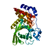
Mass: 96.063 Da / Num. of mol.: 2 / Source method: obtained synthetically / Formula: SO4
#3: Water ChemComp-HOH / water


Mass: 18.015 Da / Num. of mol.: 69 / Source method: isolated from a natural source / Formula: H2O
Has ligand of interestN

-
Experimental details

-
Experiment

ExperimentMethod: X-RAY DIFFRACTION / Number of used crystals: 1

-
Sample preparation

CrystalDensity Matthews: 2.72 Å3/Da / Density % sol: 54.77 % / Description: Rectangular
Crystal growTemperature: 293.15 K / Method: vapor diffusion, hanging drop / pH: 5.5
Details: 30% PEG 3350, 200 mM Li2SO4, 100 mM bis-tris pH 5.5

-
Data collection

DiffractionMean temperature: 310 K / Serial crystal experiment: N
Diffraction sourceSource: SYNCHROTRON / Site: CHESS / Beamline: 7B2 / Wavelength: 1.033 Å
DetectorType: DECTRIS EIGER2 S 16M / Detector: PIXEL / Date: Oct 30, 2020
RadiationMonochromator: Dual bounce W/B4C multilayer monochromator / Protocol: SINGLE WAVELENGTH / Monochromatic (M) / Laue (L): M / Scattering type: x-ray
Radiation wavelengthWavelength: 1.033 Å / Relative weight: 1
ReflectionResolution: 1.96→68.61 Å / Num. obs: 26353 / % possible obs: 99.8 % / Redundancy: 6.7 % / Biso Wilson estimate: 37 Å2 / CC1/2: 0.996 / CC star: 0.999 / Rmerge(I) obs: 0.1697 / Rpim(I) all: 0.0708 / Rrim(I) all: 0.1842 / Net I/σ(I): 7.79
Reflection shellResolution: 1.96→2.028 Å / Redundancy: 6.9 % / Rmerge(I) obs: 2.398 / Mean I/σ(I) obs: 0.63 / Num. unique obs: 2573 / CC1/2: 0.358 / CC star: 0.726 / Rpim(I) all: 0.9761 / Rrim(I) all: 2.593 / % possible all: 99.03

-
Processing

Software
NameVersionClassification
PHENIX1.19.2_4158refinement
xia2data scaling
DIMPLEv2.6.1data reduction
DIMPLEv2.6.1phasing
RefinementMethod to determine structure: MOLECULAR REPLACEMENT / Resolution: 1.96→68.61 Å / SU ML: 0.26 / Cross valid method: FREE R-VALUE / σ(F): 1.34 / Phase error: 24 / Stereochemistry target values: ML
RfactorNum. reflection% reflection
Rfree0.2057 1358 5.16 %
Rwork0.1744 --
obs0.1761 26310 99.81 %
Solvent computationShrinkage radii: 0.9 Å / VDW probe radii: 1.11 Å / Solvent model: FLAT BULK SOLVENT MODEL
Refinement stepCycle: LAST / Resolution: 1.96→68.61 Å
ProteinNucleic acidLigandSolventTotal
Num. atoms2291 0 0 69 2360
Refine LS restraints
Refine-IDTypeDev idealNumber
X-RAY DIFFRACTIONf_bond_d0.0192417
X-RAY DIFFRACTIONf_angle_d1.4043294
X-RAY DIFFRACTIONf_dihedral_angle_d6.244322
X-RAY DIFFRACTIONf_chiral_restr0.081357
X-RAY DIFFRACTIONf_plane_restr0.015429
LS refinement shell
Resolution (Å)Rfactor RfreeNum. reflection RfreeRfactor RworkNum. reflection RworkRefine-ID% reflection obs (%)
1.96-2.030.36761280.33852426X-RAY DIFFRACTION99
2.03-2.110.31531320.28582471X-RAY DIFFRACTION100
2.11-2.210.2941340.23882430X-RAY DIFFRACTION100
2.21-2.320.25381380.2222460X-RAY DIFFRACTION100
2.32-2.470.24951220.1942478X-RAY DIFFRACTION100
2.47-2.660.27361260.18382504X-RAY DIFFRACTION100
2.66-2.930.22071240.17942498X-RAY DIFFRACTION100
2.93-3.350.19541400.15832499X-RAY DIFFRACTION100
3.35-4.220.19351380.13692536X-RAY DIFFRACTION100
4.22-68.610.15981760.1572650X-RAY DIFFRACTION100

+
About Yorodumi

-
News

-
Feb 9, 2022. New format data for meta-information of EMDB entries

New format data for meta-information of EMDB entries

  • Version 3 of the EMDB header file is now the official format.
  • The previous official version 1.9 will be removed from the archive.

Related info.:EMDB header

External links:wwPDB to switch to version 3 of the EMDB data model

-
Aug 12, 2020. Covid-19 info

Covid-19 info

URL: https://pdbjlvh1.pdbj.org/emnavi/covid19.php

New page: Covid-19 featured information page in EM Navigator.

Related info.:Covid-19 info / Mar 5, 2020. Novel coronavirus structure data

+
Mar 5, 2020. Novel coronavirus structure data

Novel coronavirus structure data

Related info.:Yorodumi Speices / Aug 12, 2020. Covid-19 info

External links:COVID-19 featured content - PDBj / Molecule of the Month (242):Coronavirus Proteases

+
Jan 31, 2019. EMDB accession codes are about to change! (news from PDBe EMDB page)

EMDB accession codes are about to change! (news from PDBe EMDB page)

  • The allocation of 4 digits for EMDB accession codes will soon come to an end. Whilst these codes will remain in use, new EMDB accession codes will include an additional digit and will expand incrementally as the available range of codes is exhausted. The current 4-digit format prefixed with “EMD-” (i.e. EMD-XXXX) will advance to a 5-digit format (i.e. EMD-XXXXX), and so on. It is currently estimated that the 4-digit codes will be depleted around Spring 2019, at which point the 5-digit format will come into force.
  • The EM Navigator/Yorodumi systems omit the EMD- prefix.

Related info.:Q: What is EMD? / ID/Accession-code notation in Yorodumi/EM Navigator

External links:EMDB Accession Codes are Changing Soon! / Contact to PDBj

+
Jul 12, 2017. Major update of PDB

Major update of PDB

  • wwPDB released updated PDB data conforming to the new PDBx/mmCIF dictionary.
  • This is a major update changing the version number from 4 to 5, and with Remediation, in which all the entries are updated.
  • In this update, many items about electron microscopy experimental information are reorganized (e.g. em_software).
  • Now, EM Navigator and Yorodumi are based on the updated data.

External links:wwPDB Remediation / Enriched Model Files Conforming to OneDep Data Standards Now Available in the PDB FTP Archive

-
Yorodumi

Thousand views of thousand structures

  • Yorodumi is a browser for structure data from EMDB, PDB, SASBDB, etc.
  • This page is also the successor to EM Navigator detail page, and also detail information page/front-end page for Omokage search.
  • The word "yorodu" (or yorozu) is an old Japanese word meaning "ten thousand". "mi" (miru) is to see.

Related info.:EMDB / PDB / SASBDB / Comparison of 3 databanks / Yorodumi Search / Aug 31, 2016. New EM Navigator & Yorodumi / Yorodumi Papers / Jmol/JSmol / Function and homology information / Changes in new EM Navigator and Yorodumi

Read more