Movie
Movie Controller


+ Open data

- Basic information
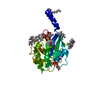
| Entry | Database: PDB / ID: 8s5c | ||||||||||||||||||||||||||||||||||||
|---|---|---|---|---|---|---|---|---|---|---|---|---|---|---|---|---|---|---|---|---|---|---|---|---|---|---|---|---|---|---|---|---|---|---|---|---|---|
| Title | Cryo-EM structure of Arf1-decorated membrane tubules | ||||||||||||||||||||||||||||||||||||
 Components Components | ADP-ribosylation factor 1 | ||||||||||||||||||||||||||||||||||||
 Keywords Keywords | TRANSPORT PROTEIN / Arf1 / protein-membrane interaction / membrane tubules / helical structure | ||||||||||||||||||||||||||||||||||||
| Function / homology |  Function and homology information Function and homology informationSynthesis of PIPs at the plasma membrane / VxPx cargo-targeting to cilium / Synthesis of PIPs at the Golgi membrane / protein localization to Golgi membrane / regulation of Golgi organization / trans-Golgi Network Vesicle Budding / organelle membrane contact site / Intra-Golgi traffic / Golgi vesicle transport / COPI-dependent Golgi-to-ER retrograde traffic ...Synthesis of PIPs at the plasma membrane / VxPx cargo-targeting to cilium / Synthesis of PIPs at the Golgi membrane / protein localization to Golgi membrane / regulation of Golgi organization / trans-Golgi Network Vesicle Budding / organelle membrane contact site / Intra-Golgi traffic / Golgi vesicle transport / COPI-dependent Golgi-to-ER retrograde traffic / COPI-mediated anterograde transport / positive regulation of mitochondrial fusion / regulation of fatty acid metabolic process / Golgi to plasma membrane transport / positive regulation of mitochondrial fission / endoplasmic reticulum to Golgi vesicle-mediated transport / vesicle-mediated transport / macroautophagy / small monomeric GTPase / intracellular protein transport / G protein activity / GTPase activity / GTP binding / Golgi apparatus / plasma membrane / cytosol / cytoplasm Similarity search - Function | ||||||||||||||||||||||||||||||||||||
| Biological species |  | ||||||||||||||||||||||||||||||||||||
| Method | ELECTRON MICROSCOPY / helical reconstruction / cryo EM / Resolution: 3.1 Å | ||||||||||||||||||||||||||||||||||||
 Authors Authors | Haupt, C. / Semchonok, D.A. / Stubbs, M.T. / Bacia, K. / Desfosses, A. / Kastritis, P.L. / Hamdi, F. | ||||||||||||||||||||||||||||||||||||
| Funding support |  Germany, 2items Germany, 2items
| ||||||||||||||||||||||||||||||||||||
 Citation Citation | Journal: Biorxiv / Year: 2026 Title: Structural basis of Arf1-driven membrane tubulation Authors: Haupt, C. / Semchonok, D.A. / Desfosses, A. / Daum, S. / Neudorf, S.S. / Hamdi, F. / Kastritis, P.L. / Stubbs, M.T. / Bacia, K. | ||||||||||||||||||||||||||||||||||||
| History |
|
- Structure visualization
| Structure viewer | Molecule: MolmilJmol/JSmol |
|---|
- Downloads & links
-Download
| PDBx/mmCIF format | 8s5c.cif.gz | 84.7 KB | Display | PDBx/mmCIF format |
|---|---|---|---|---|
| PDB format | pdb8s5c.ent.gz | 64.9 KB | Display | PDB format |
| PDBx/mmJSON format | 8s5c.json.gz | Tree view | PDBx/mmJSON format | |
| Others |  Other downloads Other downloads |
-Validation report
| Arichive directory | https://data.pdbj.org/pub/pdb/validation_reports/s5/8s5cftp://data.pdbj.org/pub/pdb/validation_reports/s5/8s5c | HTTPS FTP |
|---|
-Related structure data
| Related structure data |  19731MC  8s5dC  8s5eC M: map data used to model this data C: citing same article ( |
|---|---|
| Similar structure data |


-Links
 PDBj
PDBj


- Assembly
| Deposited unit | 
|
|---|---|
| 1 | x 205
|
-Components
| #1: Protein | Mass: 20552.438 Da / Num. of mol.: 1 Source method: isolated from a genetically manipulated source Details: A myristoyl group (derived from myristic acid) is covalently attached to the N-terminus via an amide bond. Source: (gene. exp.) Gene: ARF1, YDL192W, D1244 / Production host:  |
|---|---|
| #2: Chemical | ChemComp-MG /   Mass: 24.305 Da / Num. of mol.: 1 / Source method: obtained synthetically / Formula: Mg / Feature type: SUBJECT OF INVESTIGATION Mass: 24.305 Da / Num. of mol.: 1 / Source method: obtained synthetically / Formula: Mg / Feature type: SUBJECT OF INVESTIGATION |
| #3: Chemical | ChemComp-GSP /   Mass: 539.246 Da / Num. of mol.: 1 / Source method: obtained synthetically / Formula: C10H16N5O13P3S / Feature type: SUBJECT OF INVESTIGATION Mass: 539.246 Da / Num. of mol.: 1 / Source method: obtained synthetically / Formula: C10H16N5O13P3S / Feature type: SUBJECT OF INVESTIGATION |
| #4: Water | ChemComp-HOH /  Mass: 18.015 Da / Num. of mol.: 2 / Source method: isolated from a natural source / Formula: H2O Mass: 18.015 Da / Num. of mol.: 2 / Source method: isolated from a natural source / Formula: H2O |
| Has ligand of interest | Y |
| Has protein modification | N |
-Experimental details
-Experiment
| Experiment | Method: ELECTRON MICROSCOPY |
|---|---|
| EM experiment | Aggregation state: HELICAL ARRAY / 3D reconstruction method: helical reconstruction |
- Sample preparation
| Component | Name: Arf1 coated membrane tubules / Type: COMPLEX / Entity ID: #1 / Source: RECOMBINANT | ||||||||||||||||||||
|---|---|---|---|---|---|---|---|---|---|---|---|---|---|---|---|---|---|---|---|---|---|
| Molecular weight | Experimental value: NO | ||||||||||||||||||||
| Source (natural) | Organism: | ||||||||||||||||||||
| Source (recombinant) | Organism: | ||||||||||||||||||||
| Buffer solution | pH: 6.8 | ||||||||||||||||||||
| Buffer component |
| ||||||||||||||||||||
| Specimen | Embedding applied: NO / Shadowing applied: NO / Staining applied: NO / Vitrification applied: YES | ||||||||||||||||||||
| Specimen support | Grid material: COPPER / Grid mesh size: 200 divisions/in. / Grid type: Quantifoil R2/1 | ||||||||||||||||||||
| Vitrification | Instrument: LEICA EM GP / Cryogen name: ETHANE / Humidity: 80 % / Chamber temperature: 277 K |
- Electron microscopy imaging
| Microscopy | Model: TFS GLACIOS |
|---|---|
| Electron gun | Electron source:  FIELD EMISSION GUN / Accelerating voltage: 200 kV / Illumination mode: OTHER FIELD EMISSION GUN / Accelerating voltage: 200 kV / Illumination mode: OTHER |
| Electron lens | Mode: BRIGHT FIELD / Nominal magnification: 92000 X / Nominal defocus max: 2800 nm / Nominal defocus min: 600 nm / Cs: 2.7 mm / C2 aperture diameter: 70 µm / Alignment procedure: COMA FREE |
| Specimen holder | Cryogen: NITROGEN / Specimen holder model: OTHER |
| Image recording | Electron dose: 21 e/Å2 / Film or detector model: FEI FALCON IV (4k x 4k) / Num. of grids imaged: 1 / Num. of real images: 12483 |
| Image scans | Width: 4096 / Height: 4096 |
- Processing
| EM software |
| ||||||||||||||||||||||||||||||||||||
|---|---|---|---|---|---|---|---|---|---|---|---|---|---|---|---|---|---|---|---|---|---|---|---|---|---|---|---|---|---|---|---|---|---|---|---|---|---|
| CTF correction | Type: PHASE FLIPPING AND AMPLITUDE CORRECTION | ||||||||||||||||||||||||||||||||||||
| Helical symmerty | Angular rotation/subunit: 23.27 ° / Axial rise/subunit: 4.328 Å / Axial symmetry: C2 | ||||||||||||||||||||||||||||||||||||
| Particle selection | Num. of particles selected: 1489051 | ||||||||||||||||||||||||||||||||||||
| 3D reconstruction | Resolution: 3.1 Å / Resolution method: FSC 0.143 CUT-OFF / Num. of particles: 127727 / Algorithm: FOURIER SPACE / Num. of class averages: 1 / Symmetry type: HELICAL | ||||||||||||||||||||||||||||||||||||
| Atomic model building | Protocol: OTHER / Space: REAL | ||||||||||||||||||||||||||||||||||||
| Atomic model building | PDB-ID: 2ksq Pdb chain-ID: A / Accession code: 2ksq / Chain residue range: 16-178 / Pdb chain residue range: 16-178 / Source name: PDB / Type: experimental model |