Movie
Movie Controller


+ Open data

- Basic information
| Entry |  | |||||||||
|---|---|---|---|---|---|---|---|---|---|---|
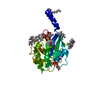
| Title | Cryo-EM structure of Arf1-decorated membrane tubules | |||||||||
 Map data Map data | map_sharp | |||||||||
 Sample Sample |
| |||||||||
 Keywords Keywords | Arf1 / protein-membrane interaction / membrane tubules / helical structure / TRANSPORT PROTEIN | |||||||||
| Function / homology |  Function and homology information Function and homology informationSynthesis of PIPs at the plasma membrane / VxPx cargo-targeting to cilium / Synthesis of PIPs at the Golgi membrane / protein localization to Golgi membrane / regulation of Golgi organization / trans-Golgi Network Vesicle Budding / organelle membrane contact site / Intra-Golgi traffic / Golgi vesicle transport / COPI-dependent Golgi-to-ER retrograde traffic ...Synthesis of PIPs at the plasma membrane / VxPx cargo-targeting to cilium / Synthesis of PIPs at the Golgi membrane / protein localization to Golgi membrane / regulation of Golgi organization / trans-Golgi Network Vesicle Budding / organelle membrane contact site / Intra-Golgi traffic / Golgi vesicle transport / COPI-dependent Golgi-to-ER retrograde traffic / COPI-mediated anterograde transport / positive regulation of mitochondrial fusion / regulation of fatty acid metabolic process / Golgi to plasma membrane transport / positive regulation of mitochondrial fission / endoplasmic reticulum to Golgi vesicle-mediated transport / vesicle-mediated transport / small monomeric GTPase / macroautophagy / intracellular protein transport / G protein activity / GTPase activity / GTP binding / Golgi apparatus / plasma membrane / cytoplasm / cytosol Similarity search - Function | |||||||||
| Biological species |  | |||||||||
| Method | helical reconstruction / cryo EM / Resolution: 3.79 Å | |||||||||
 Authors Authors | Haupt C / Semchonok DA / Stubbs MT / Bacia K / Desfosses A / Kastritis PL / Hamdi F | |||||||||
| Funding support |  Germany, 2 items Germany, 2 items
| |||||||||
 Citation Citation | Journal: Biorxiv / Year: 2026 Title: Structural basis of Arf1-driven membrane tubulation Authors: Haupt C / Semchonok DA / Desfosses A / Daum S / Neudorf SS / Hamdi F / Kastritis PL / Stubbs MT / Bacia K | |||||||||
| History |
|
- Structure visualization
| Supplemental images |
|---|

- Downloads & links
-EMDB archive
| Map data | emd_19732.map.gz | 203.7 MB | EMDB map data format | |
|---|---|---|---|---|
| Header (meta data) | emd-19732-v30.xmlemd-19732.xml | 30.3 KB 30.3 KB | Display Display | EMDB header |
| FSC (resolution estimation) | emd_19732_fsc.xml | 12.6 KB | Display | FSC data file |
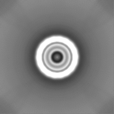
| Images |  emd_19732.png emd_19732.png | 79.1 KB | ||
| Masks | emd_19732_msk_1.map | 216 MB | Mask map | |
| Filedesc metadata | emd-19732.cif.gz | 7.2 KB | ||
| Others | emd_19732_additional_1.map.gzemd_19732_additional_2.map.gzemd_19732_additional_3.map.gzemd_19732_half_map_1.map.gzemd_19732_half_map_2.map.gz | 31.5 MB 31.6 MB 108.5 MB 200.6 MB 200.6 MB | ||
| Archive directory |  http://ftp.pdbj.org/pub/emdb/structures/EMD-19732ftp://ftp.pdbj.org/pub/emdb/structures/EMD-19732 http://ftp.pdbj.org/pub/emdb/structures/EMD-19732ftp://ftp.pdbj.org/pub/emdb/structures/EMD-19732 | HTTPS FTP |
-Related structure data
| Related structure data |  8s5dMC  8s5cC  8s5eC M: atomic model generated by this map C: citing same article ( |
|---|---|
| Similar structure data |


-Links
| EMDB pages | EMDB (EBI/PDBe) / EMDataResource |
|---|---|
| Related items in Molecule of the Month |


-Map
| File | Download / File: emd_19732.map.gz / Format: CCP4 / Size: 216 MB / Type: IMAGE STORED AS FLOATING POINT NUMBER (4 BYTES) | ||||||||||||||||||||||||||||||||||||
|---|---|---|---|---|---|---|---|---|---|---|---|---|---|---|---|---|---|---|---|---|---|---|---|---|---|---|---|---|---|---|---|---|---|---|---|---|---|
| Annotation | map_sharp | ||||||||||||||||||||||||||||||||||||
| Projections & slices | Image control
Images are generated by Spider. | ||||||||||||||||||||||||||||||||||||
| Voxel size | X=Y=Z: 1.53041 Å | ||||||||||||||||||||||||||||||||||||
| Density |
| ||||||||||||||||||||||||||||||||||||
| Symmetry | Space group: 1 | ||||||||||||||||||||||||||||||||||||
| Details | EMDB XML:
|
 Z (Sec.)
 Y (Row.)
 X (Col.)


-Supplemental data
-Mask #1
| File | emd_19732_msk_1.map | ||||||||||||
|---|---|---|---|---|---|---|---|---|---|---|---|---|---|
| Projections & Slices |
| ||||||||||||
| Density Histograms |


-Additional map: map sym
| File | emd_19732_additional_1.map | ||||||||||||
|---|---|---|---|---|---|---|---|---|---|---|---|---|---|
| Annotation | map_sym | ||||||||||||
| Projections & Slices |
| ||||||||||||
| Density Histograms |


-Additional map: map sym sharp
| File | emd_19732_additional_2.map | ||||||||||||
|---|---|---|---|---|---|---|---|---|---|---|---|---|---|
| Annotation | map_sym_sharp | ||||||||||||
| Projections & Slices |
| ||||||||||||
| Density Histograms |


-Additional map: map
| File | emd_19732_additional_3.map | ||||||||||||
|---|---|---|---|---|---|---|---|---|---|---|---|---|---|
| Annotation | map | ||||||||||||
| Projections & Slices |
| ||||||||||||
| Density Histograms |


-Half map: B
| File | emd_19732_half_map_1.map | ||||||||||||
|---|---|---|---|---|---|---|---|---|---|---|---|---|---|
| Annotation | B | ||||||||||||
| Projections & Slices |
| ||||||||||||
| Density Histograms |


-Half map: A
| File | emd_19732_half_map_2.map | ||||||||||||
|---|---|---|---|---|---|---|---|---|---|---|---|---|---|
| Annotation | A | ||||||||||||
| Projections & Slices |
| ||||||||||||
| Density Histograms |


- Sample components
-Entire : Arf1 coated membrane tubules
| Entire | Name: Arf1 coated membrane tubules |
|---|---|
| Components |
|
-Supramolecule #1: Arf1 coated membrane tubules
| Supramolecule | Name: Arf1 coated membrane tubules / type: complex / ID: 1 / Parent: 0 / Macromolecule list: #1 |
|---|---|
| Source (natural) | Organism: |
-Macromolecule #1: ADP-ribosylation factor 1
| Macromolecule | Name: ADP-ribosylation factor 1 / type: protein_or_peptide / ID: 1 Details: A myristoyl group (derived from myristic acid) is covalently attached to the N-terminus via an amide bond. Number of copies: 1 / Enantiomer: LEVO / EC number: small monomeric GTPase |
|---|---|
| Source (natural) | Organism: |
| Molecular weight | Theoretical: 20.552438 KDa |
| Recombinant expression | Organism:  |
| Sequence | String: MGLFASKLFS NLFGNKEMRI LMVGLDGAGK TTVLYKLKLG EVITTIPTIG FNVETVQYKN ISFTVWDVGG QDRIRSLWRH YYRNTEGVI FVVDSNDRSR IGEAREVMQR MLNEDELRNA AWLVFANKQD LPEAMSAAEI TEKLGLHSIR NRPWFIQATC A TSGEGLYE GLEWLSNSLK NST UniProtKB: ADP-ribosylation factor 1 |
-Macromolecule #2: MAGNESIUM ION
| Macromolecule | Name: MAGNESIUM ION / type: ligand / ID: 2 / Number of copies: 1 / Formula: MG |
|---|---|
| Molecular weight | Theoretical: 24.305 Da |
-Macromolecule #3: 5'-GUANOSINE-DIPHOSPHATE-MONOTHIOPHOSPHATE
| Macromolecule | Name: 5'-GUANOSINE-DIPHOSPHATE-MONOTHIOPHOSPHATE / type: ligand / ID: 3 / Number of copies: 1 / Formula: GSP |
|---|---|
| Molecular weight | Theoretical: 539.246 Da |
| Chemical component information |  ChemComp-GSP: |
-Macromolecule #4: water
| Macromolecule | Name: water / type: ligand / ID: 4 / Number of copies: 2 / Formula: HOH |
|---|---|
| Molecular weight | Theoretical: 18.015 Da |
| Chemical component information |  ChemComp-HOH: |
-Experimental details
-Structure determination
| Method | cryo EM |
|---|---|
 Processing Processing | helical reconstruction |
| Aggregation state | helical array |
-Sample preparation
| Buffer | pH: 6.8 Component:
| ||||||||||||
|---|---|---|---|---|---|---|---|---|---|---|---|---|---|
| Grid | Model: Quantifoil R2/1 / Material: COPPER / Mesh: 200 / Support film - Material: CARBON / Support film - topology: HOLEY ARRAY / Pretreatment - Type: PLASMA CLEANING / Pretreatment - Time: 25 sec. / Pretreatment - Atmosphere: AIR | ||||||||||||
| Vitrification | Cryogen name: ETHANE / Chamber humidity: 80 % / Chamber temperature: 277 K / Instrument: LEICA EM GP |
- Electron microscopy
| Microscope | TFS GLACIOS |
|---|---|
| Image recording | Film or detector model: FEI FALCON IV (4k x 4k) / Digitization - Dimensions - Width: 4096 pixel / Digitization - Dimensions - Height: 4096 pixel / Number grids imaged: 1 / Number real images: 12483 / Average electron dose: 21.0 e/Å2 |
| Electron beam | Acceleration voltage: 200 kV / Electron source:  FIELD EMISSION GUN FIELD EMISSION GUN |
| Electron optics | C2 aperture diameter: 70.0 µm / Illumination mode: OTHER / Imaging mode: BRIGHT FIELD / Cs: 2.7 mm / Nominal defocus max: 2.8000000000000003 µm / Nominal defocus min: 0.6 µm / Nominal magnification: 92000 |
| Sample stage | Specimen holder model: OTHER / Cooling holder cryogen: NITROGEN |