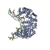
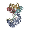
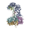
Journal: Nat Commun / Year: 2025 Title: A FAN1 point mutation associated with accelerated Huntington's disease progression alters its PCNA-mediated assembly on DNA. Authors: Jonas Aretz / Gayathri Jeyasankar / Anna Salerno-Kochan / Maren Thomsen / Gabriel Thieulin-Pardo / Tasir Haque / Edith Monteagudo / Dan Felsenfeld / Michael Finley / Thomas F Vogt / Julien ...Authors: Jonas Aretz / Gayathri Jeyasankar / Anna Salerno-Kochan / Maren Thomsen / Gabriel Thieulin-Pardo / Tasir Haque / Edith Monteagudo / Dan Felsenfeld / Michael Finley / Thomas F Vogt / Julien Boudet / Brinda C Prasad / Abstract: FAN1 is an endo- and exo-nuclease involved in DNA and interstrand crosslink repair. Genome-wide association studies of people with Huntington's disease revealed a strong association between the FAN1 ...FAN1 is an endo- and exo-nuclease involved in DNA and interstrand crosslink repair. Genome-wide association studies of people with Huntington's disease revealed a strong association between the FAN1 R507H mutation and early disease onset, however the underlying mechanism(s) remains unclear. FAN1 has previously been implicated in modulating triplet repeat expansion in a PCNA dependent manner. To examine the role of PCNA on FAN1 activation, we solved the cryo-EM structures of a PCNA-FAN1-DNA complex. Our findings reveal that the FAN1 R507 residue directly interacts with PCNA D232. Biophysical interaction studies demonstrated that FAN1 enhances the binding affinity of PCNA for DNA, a synergistic effect disrupted in mutants carrying the R507H mutation. In contrast, PCNA does not affect the affinity of FAN1 for DNA but does modulate FAN1 activity upon ternary complex formation. The weakened and functionally altered FAN1 R507H-PCNA-DNA complex may partly impair the FAN1-mediated repair of CAG extrahelical extrusions, providing a potential explanation for the mutation's role in accelerating disease progression.
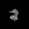
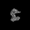
A: Fanconi-associated nuclease 1 B: DNA (5'-D(*AP*AP*CP*AP*CP*GP*CP*CP*TP*AP*GP*AP*CP*TP*CP*CP*TP*CP*A)-3') C: DNA (5'-D(P*GP*AP*GP*GP*CP*GP*TP*G)-3') D: DNA (5'-D(*TP*TP*TP*GP*AP*GP*GP*AP*GP*TP*CP*TP*T)-3') hetero molecules
In the structure databanks used in Yorodumi, some data are registered as the other names, "COVID-19 virus" and "2019-nCoV". Here are the details of the virus and the list of structure data.
Jan 31, 2019. EMDB accession codes are about to change! (news from PDBe EMDB page)
EMDB accession codes are about to change! (news from PDBe EMDB page)
The allocation of 4 digits for EMDB accession codes will soon come to an end. Whilst these codes will remain in use, new EMDB accession codes will include an additional digit and will expand incrementally as the available range of codes is exhausted. The current 4-digit format prefixed with “EMD-” (i.e. EMD-XXXX) will advance to a 5-digit format (i.e. EMD-XXXXX), and so on. It is currently estimated that the 4-digit codes will be depleted around Spring 2019, at which point the 5-digit format will come into force.
The EM Navigator/Yorodumi systems omit the EMD- prefix.
Related info.:Q: What is EMD? / ID/Accession-code notation in Yorodumi/EM Navigator
Yorodumi is a browser for structure data from EMDB, PDB, SASBDB, etc.
This page is also the successor to EM Navigator detail page, and also detail information page/front-end page for Omokage search.
The word "yorodu" (or yorozu) is an old Japanese word meaning "ten thousand". "mi" (miru) is to see.
Related info.:EMDB / PDB / SASBDB / Comparison of 3 databanks / Yorodumi Search / Aug 31, 2016. New EM Navigator & Yorodumi / Yorodumi Papers / Jmol/JSmol / Function and homology information / Changes in new EM Navigator and Yorodumi
 Movie
Movie Controller

 Yorodumi
 Open data
 Basic information
 Components
 Keywords
 Function and homology information
 Homo sapiens (human)
 X-RAY DIFFRACTION /
  Authors
 Citation

 Structure visualization
 Downloads & links
 Other downloads
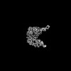

 PDBj
PDBj


 Assembly

 Spodoptera frugiperda (fall armyworm)


 Mass: 40.078 Da / Num. of mol.: 2 / Source method: obtained synthetically / Formula: Ca
 Mass: 35.453 Da / Num. of mol.: 1 / Source method: isolated from a natural source / Formula: Cl
 Sample preparation
 / Beamline: 08ID-1 / Wavelength: 0.9537 Å
 Processing