 Movie
Movie Controller


+ Open data

- Basic information
| Entry | Database: PDB / ID: 8rxc | |||||||||||||||||||||||||||||||||||||||||||||
|---|---|---|---|---|---|---|---|---|---|---|---|---|---|---|---|---|---|---|---|---|---|---|---|---|---|---|---|---|---|---|---|---|---|---|---|---|---|---|---|---|---|---|---|---|---|---|
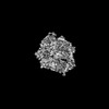
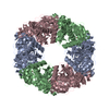
| Title | RadA helicase from Streptococcus pneumoniae coordinating dsDNA | |||||||||||||||||||||||||||||||||||||||||||||
 Components Components |
| |||||||||||||||||||||||||||||||||||||||||||||
 Keywords Keywords | DNA BINDING PROTEIN / Helicase Translocase natural transformation | |||||||||||||||||||||||||||||||||||||||||||||
| Function / homology |  Function and homology information Function and homology informationrecombinational repair / ATP-dependent DNA damage sensor activity / damaged DNA binding / hydrolase activity / zinc ion binding / ATP binding / cytosol Similarity search - Function | |||||||||||||||||||||||||||||||||||||||||||||
| Biological species |   Streptococcus pneumoniae (bacteria) Streptococcus pneumoniae (bacteria)synthetic construct (others) | |||||||||||||||||||||||||||||||||||||||||||||
| Method | ELECTRON MICROSCOPY / single particle reconstruction / cryo EM / Resolution: 3.15 Å | |||||||||||||||||||||||||||||||||||||||||||||
 Authors Authors | Talachia Rosa, L. / Fronzes, R. | |||||||||||||||||||||||||||||||||||||||||||||
| Funding support | European Union, 1items
| |||||||||||||||||||||||||||||||||||||||||||||
 Citation Citation | Journal: EMBO J / Year: 2024 Title: Structural insights into the mechanism of DNA branch migration during homologous recombination in bacteria. Authors: Leonardo Talachia Rosa / Émeline Vernhes / Anne-Lise Soulet / Patrice Polard / Rémi Fronzes /  Abstract: Some DNA helicases play central and specific roles in genome maintenance and plasticity through their branch migration activity in different pathways of homologous recombination. RadA is a highly ...Some DNA helicases play central and specific roles in genome maintenance and plasticity through their branch migration activity in different pathways of homologous recombination. RadA is a highly conserved bacterial helicase involved in DNA repair throughout all bacterial species. In Gram-positive Firmicutes, it also has a role in natural transformation, while in Gram-negative bacteria, ComM is the canonical transformation-specific helicase. Both RadA and ComM helicases form hexameric rings and use ATP hydrolysis as an energy source to propel themselves along DNA. In this study, we present the cryoEM structures of RadA and ComM interacting with DNA and ATP analogs. These structures reveal important molecular interactions that couple ATP hydrolysis and DNA binding in RadA, as well as the role of the Lon protease-like domain, shared by RadA and ComM, in this process. Taken together, these results provide new molecular insights into the mechanisms of DNA branch migration in different pathways of homologous recombination. | |||||||||||||||||||||||||||||||||||||||||||||
| History |
|
- Structure visualization
| Structure viewer | Molecule: MolmilJmol/JSmol |
|---|
- Downloads & links
-Download
| PDBx/mmCIF format | 8rxc.cif.gz | 711.5 KB | Display | PDBx/mmCIF format |
|---|---|---|---|---|
| PDB format | pdb8rxc.ent.gz | Display | PDB format | |
| PDBx/mmJSON format | 8rxc.json.gz | Tree view | PDBx/mmJSON format | |
| Others |  Other downloads Other downloads |
-Validation report
| Arichive directory | https://data.pdbj.org/pub/pdb/validation_reports/rx/8rxcftp://data.pdbj.org/pub/pdb/validation_reports/rx/8rxc | HTTPS FTP |
|---|
-Related structure data
| Related structure data | 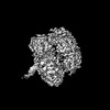 19573MC  8rxdC  8rxkC  8rxsC 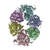 8rxtC M: map data used to model this data C: citing same article ( |
|---|---|
| Similar structure data |


-Links
 PDBj
PDBj


- Assembly
| Deposited unit | 
|
|---|---|
| 1 |
|
-Components
| #1: Protein | Mass: 51526.953 Da / Num. of mol.: 6 Source method: isolated from a genetically manipulated source Source: (gene. exp.) Streptococcus pneumoniae (bacteria)Gene: radA, A5N45_07085, GM529_10050, GM532_03345, GM536_10290, GM537_09620, GM538_09765, GM539_03745, GM543_09545, GM544_10415, GM546_02225 Production host: #2: DNA chain | | Mass: 9080.827 Da / Num. of mol.: 1 / Source method: obtained synthetically / Source: (synth.) synthetic construct (others) #3: DNA chain | | Mass: 36098.395 Da / Num. of mol.: 1 / Source method: obtained synthetically / Source: (synth.) synthetic construct (others) #4: Chemical | ChemComp-AGS /   Mass: 523.247 Da / Num. of mol.: 5 / Source method: obtained synthetically / Formula: C10H16N5O12P3S / Feature type: SUBJECT OF INVESTIGATION / Comment: ATP-gamma-S, energy-carrying molecule analogue*YM Mass: 523.247 Da / Num. of mol.: 5 / Source method: obtained synthetically / Formula: C10H16N5O12P3S / Feature type: SUBJECT OF INVESTIGATION / Comment: ATP-gamma-S, energy-carrying molecule analogue*YM#5: Chemical | ChemComp-MG /   Mass: 24.305 Da / Num. of mol.: 5 / Source method: obtained synthetically / Formula: Mg / Feature type: SUBJECT OF INVESTIGATION Mass: 24.305 Da / Num. of mol.: 5 / Source method: obtained synthetically / Formula: Mg / Feature type: SUBJECT OF INVESTIGATIONHas ligand of interest | Y | Has protein modification | N | |
|---|
-Experimental details
-Experiment
| Experiment | Method: ELECTRON MICROSCOPY |
|---|---|
| EM experiment | Aggregation state: PARTICLE / 3D reconstruction method: single particle reconstruction |
- Sample preparation
| Component | Name: RadA helicase from Streptococcus pneumoniae coordinating dsDNA Type: COMPLEX / Entity ID: #1-#3 / Source: RECOMBINANT |
|---|---|
| Source (natural) | Organism: Streptococcus pneumoniae (bacteria) |
| Source (recombinant) | Organism: |
| Buffer solution | pH: 8 |
| Specimen | Conc.: 0.35 mg/ml / Embedding applied: NO / Shadowing applied: NO / Staining applied: NO / Vitrification applied: YES / Details: 0.35 mg/ml protein and 100 uM of hybrid DNA |
| Vitrification | Instrument: FEI VITROBOT MARK III / Cryogen name: ETHANE / Humidity: 100 % |
- Electron microscopy imaging
| Experimental equipment |  Model: Titan Krios / Image courtesy: FEI Company |
|---|---|
| Microscopy | Model: FEI TITAN KRIOS |
| Electron gun | Electron source:  FIELD EMISSION GUN / Accelerating voltage: 300 kV / Illumination mode: FLOOD BEAM FIELD EMISSION GUN / Accelerating voltage: 300 kV / Illumination mode: FLOOD BEAM |
| Electron lens | Mode: BRIGHT FIELD / Nominal defocus max: 2500 nm / Nominal defocus min: 500 nm |
| Image recording | Electron dose: 54.04 e/Å2 / Detector mode: COUNTING / Film or detector model: FEI FALCON III (4k x 4k) |
- Processing
| EM software | Name: PHENIX / Category: model refinement | ||||||||||||||||||||||||
|---|---|---|---|---|---|---|---|---|---|---|---|---|---|---|---|---|---|---|---|---|---|---|---|---|---|
| CTF correction | Type: PHASE FLIPPING AND AMPLITUDE CORRECTION | ||||||||||||||||||||||||
| 3D reconstruction | Resolution: 3.15 Å / Resolution method: FSC 0.143 CUT-OFF / Num. of particles: 303651 / Symmetry type: POINT | ||||||||||||||||||||||||
| Refine LS restraints |
|
