 Movie
Movie Controller

[English] 日本語
 Yorodumi
- PDB-8rss: Crystal structure of marine actinobacteria clade rhodopsin (MAR) ... -
+ Open data

- Basic information
| Entry | Database: PDB / ID: 8rss | ||||||
|---|---|---|---|---|---|---|---|
| Title | Crystal structure of marine actinobacteria clade rhodopsin (MAR) in the O* state | ||||||
 Components Components | Bacteriorhodopsin | ||||||
 Keywords Keywords | MEMBRANE PROTEIN / Mac / MacR / MAR / proteorhodopsin / PR / xanthorodopsin / XR / xanthorhodopsin | ||||||
| Function / homology | Bacteriorhodopsin-like protein / Archaeal/bacterial/fungal rhodopsins / Bacteriorhodopsin-like protein / membrane / EICOSANE / OLEIC ACID / RETINAL / Bacteriorhodopsin Function and homology information Function and homology information | ||||||
| Biological species |  Candidatus Actinomarina minuta (bacteria)marine Actinobacteria clade (bacteria) Candidatus Actinomarina minuta (bacteria)marine Actinobacteria clade (bacteria) | ||||||
| Method |  X-RAY DIFFRACTION / SYNCHROTRON / MOLECULAR REPLACEMENT / Resolution: 1.41 Å X-RAY DIFFRACTION / SYNCHROTRON / MOLECULAR REPLACEMENT / Resolution: 1.41 Å | ||||||
 Authors Authors | Bukhdruker, S. / Kovalev, K. / Astashkin, R. / Gordeliy, V. | ||||||
| Funding support |  France, 1items France, 1items
| ||||||
 Citation Citation | Journal: Sci Adv / Year: 2025 Title: Proteorhodopsin insights into the molecular mechanism of vectorial proton transport. Authors: Bukhdruker, S. / Gushchin, I. / Shevchenko, V. / Kovalev, K. / Polovinkin, V. / Tsybrov, F. / Astashkin, R. / Alekseev, A. / Mikhaylov, A. / Bukhalovich, S. / Bratanov, D. / Ryzhykau, Y. / ...Authors: Bukhdruker, S. / Gushchin, I. / Shevchenko, V. / Kovalev, K. / Polovinkin, V. / Tsybrov, F. / Astashkin, R. / Alekseev, A. / Mikhaylov, A. / Bukhalovich, S. / Bratanov, D. / Ryzhykau, Y. / Kuklina, D. / Caramello, N. / Rokitskaya, T. / Antonenko, Y. / Rulev, M. / Stoev, C. / Zabelskii, D. / Round, E. / Rogachev, A. / Borshchevskiy, V. / Ghai, R. / Bourenkov, G. / Zeghouf, M. / Cherfils, J. / Engelhard, M. / Chizhov, I. / Rodriguez-Valera, F. / Bamberg, E. / Gordeliy, V. | ||||||
| History |
|
- Structure visualization
| Structure viewer | Molecule: MolmilJmol/JSmol |
|---|
- Downloads & links
-Download
| PDBx/mmCIF format | 8rss.cif.gz | 142.1 KB | Display | PDBx/mmCIF format |
|---|---|---|---|---|
| PDB format | pdb8rss.ent.gz | 90.8 KB | Display | PDB format |
| PDBx/mmJSON format | 8rss.json.gz | Tree view | PDBx/mmJSON format | |
| Others |  Other downloads Other downloads |
-Validation report
| Summary document | 8rss_validation.pdf.gz | 1.7 MB | Display | wwPDB validaton report |
|---|---|---|---|---|
| Full document | 8rss_full_validation.pdf.gz | 1.7 MB | Display | |
| Data in XML | 8rss_validation.xml.gz | 15 KB | Display | |
| Data in CIF | 8rss_validation.cif.gz | 20.4 KB | Display | |
| Arichive directory | https://data.pdbj.org/pub/pdb/validation_reports/rs/8rssftp://data.pdbj.org/pub/pdb/validation_reports/rs/8rss | HTTPS FTP |
-Related structure data
| Related structure data | 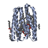 7avnC 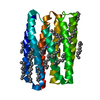 7avpC  8rsoC  8rspC  8rsqC  8rsrC 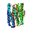 9g15C  9g16C C: citing same article ( |
|---|---|
| Similar structure data |
-Links
 PDBj
PDBj


- Assembly
| Deposited unit | 
| ||||||||||||
|---|---|---|---|---|---|---|---|---|---|---|---|---|---|
| 1 |
| ||||||||||||
| Unit cell |
| ||||||||||||
| Components on special symmetry positions |
|
-Components
| #1: Protein | Mass: 24277.076 Da / Num. of mol.: 1 Source method: isolated from a genetically manipulated source Source: (gene. exp.) Candidatus Actinomarina minuta (bacteria)Production host: | ||||||||||
|---|---|---|---|---|---|---|---|---|---|---|---|
| #2: Chemical |   Mass: 282.461 Da / Num. of mol.: 3 / Source method: obtained synthetically / Formula: C18H34O2 Mass: 282.461 Da / Num. of mol.: 3 / Source method: obtained synthetically / Formula: C18H34O2#3: Chemical | ChemComp-LFA /   Mass: 282.547 Da / Num. of mol.: 11 / Source method: obtained synthetically / Formula: C20H42 Mass: 282.547 Da / Num. of mol.: 11 / Source method: obtained synthetically / Formula: C20H42#4: Chemical | ChemComp-RET / |   Mass: 284.436 Da / Num. of mol.: 1 Mass: 284.436 Da / Num. of mol.: 1Source method: isolated from a genetically manipulated source Formula: C20H28O / Source: (gene. exp.) marine Actinobacteria clade (bacteria) / Production host: #5: Water | ChemComp-HOH / |  Mass: 18.015 Da / Num. of mol.: 147 / Source method: isolated from a natural source / Formula: H2O Mass: 18.015 Da / Num. of mol.: 147 / Source method: isolated from a natural source / Formula: H2OHas ligand of interest | Y | Has protein modification | Y | |
-Experimental details
-Experiment
| Experiment | Method: X-RAY DIFFRACTION / Number of used crystals: 1 |
|---|
- Sample preparation
| Crystal | Density Matthews: 2.45 Å3/Da / Density % sol: 49.8 % |
|---|---|
| Crystal grow | Temperature: 293 K / Method: lipidic cubic phase / pH: 8.4 / Details: 2.6 M Ammonium Phosphate buffer |
-Data collection
| Diffraction | Mean temperature: 100 K / Serial crystal experiment: N | |||||||||||||||||||||
|---|---|---|---|---|---|---|---|---|---|---|---|---|---|---|---|---|---|---|---|---|---|---|
| Diffraction source | Source: SYNCHROTRON / Site: ESRF / Beamline: ID23-1 / Wavelength: 0.8856 Å | |||||||||||||||||||||
| Detector | Type: DECTRIS EIGER2 X 16M / Detector: PIXEL / Date: Jan 20, 2022 | |||||||||||||||||||||
| Radiation | Protocol: SINGLE WAVELENGTH / Monochromatic (M) / Laue (L): M / Scattering type: x-ray | |||||||||||||||||||||
| Radiation wavelength | Wavelength: 0.8856 Å / Relative weight: 1 | |||||||||||||||||||||
| Reflection | Resolution: 1.41→40.17 Å / Num. obs: 27492 / % possible obs: 88.8 % / Redundancy: 2.5 % / Biso Wilson estimate: 14.12 Å2 / CC1/2: 0.998 / Rpim(I) all: 0.047 / Net I/σ(I): 7 | |||||||||||||||||||||
| Reflection shell | Diffraction-ID: 1
|
- Processing
| Software |
| |||||||||||||||||||||||||||||||||||||||||||||||||||||||||||||||||||||||||||||
|---|---|---|---|---|---|---|---|---|---|---|---|---|---|---|---|---|---|---|---|---|---|---|---|---|---|---|---|---|---|---|---|---|---|---|---|---|---|---|---|---|---|---|---|---|---|---|---|---|---|---|---|---|---|---|---|---|---|---|---|---|---|---|---|---|---|---|---|---|---|---|---|---|---|---|---|---|---|---|
| Refinement | Method to determine structure: MOLECULAR REPLACEMENT / Resolution: 1.41→40.17 Å / SU ML: 0.1103 / Cross valid method: FREE R-VALUE / σ(F): 1.36 / Phase error: 24.4139 Stereochemistry target values: GeoStd + Monomer Library + CDL v1.2
| |||||||||||||||||||||||||||||||||||||||||||||||||||||||||||||||||||||||||||||
| Solvent computation | Shrinkage radii: 0.9 Å / VDW probe radii: 1.1 Å / Solvent model: FLAT BULK SOLVENT MODEL | |||||||||||||||||||||||||||||||||||||||||||||||||||||||||||||||||||||||||||||
| Displacement parameters | Biso mean: 20.08 Å2 | |||||||||||||||||||||||||||||||||||||||||||||||||||||||||||||||||||||||||||||
| Refinement step | Cycle: LAST / Resolution: 1.41→40.17 Å
| |||||||||||||||||||||||||||||||||||||||||||||||||||||||||||||||||||||||||||||
| Refine LS restraints |
| |||||||||||||||||||||||||||||||||||||||||||||||||||||||||||||||||||||||||||||
| LS refinement shell |
|
