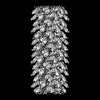
 Movie
Movie Controller


+ Open data

- Basic information
| Entry | Database: PDB / ID: 8rqb | ||||||
|---|---|---|---|---|---|---|---|
| Title | Cryo-EM structure of mouse heavy-chain apoferritin | ||||||
 Components Components | Ferritin heavy chain, N-terminally processed | ||||||
 Keywords Keywords | METAL BINDING PROTEIN / apoferritin / mouse / heavy-chain / high resolution | ||||||
| Function / homology |  Function and homology information Function and homology informationIron uptake and transport / Golgi Associated Vesicle Biogenesis / ferroxidase / negative regulation of ferroptosis / autolysosome / ferroxidase activity / negative regulation of fibroblast proliferation / endocytic vesicle lumen / Neutrophil degranulation / ferric iron binding ...Iron uptake and transport / Golgi Associated Vesicle Biogenesis / ferroxidase / negative regulation of ferroptosis / autolysosome / ferroxidase activity / negative regulation of fibroblast proliferation / endocytic vesicle lumen / Neutrophil degranulation / ferric iron binding / autophagosome / iron ion transport / ferrous iron binding / intracellular iron ion homeostasis / immune response / iron ion binding / negative regulation of cell population proliferation / mitochondrion / extracellular region / membrane / identical protein binding / cytoplasm / cytosol Similarity search - Function | ||||||
| Biological species |  | ||||||
| Method | ELECTRON MICROSCOPY / single particle reconstruction / cryo EM / Resolution: 1.09 Å | ||||||
 Authors Authors | Nazarov, S. / Myasnikov, A. / Stahlberg, H. | ||||||
| Funding support |  Switzerland, 1items Switzerland, 1items
| ||||||
 Citation Citation | Journal: Nat Commun / Year: 2024 Title: Low-dose cryo-electron ptychography of proteins at sub-nanometer resolution. Authors: Berk Küçükoğlu / Inayathulla Mohammed / Ricardo C Guerrero-Ferreira / Stephanie M Ribet / Georgios Varnavides / Max Leo Leidl / Kelvin Lau / Sergey Nazarov / Alexander Myasnikov / ...Authors: Berk Küçükoğlu / Inayathulla Mohammed / Ricardo C Guerrero-Ferreira / Stephanie M Ribet / Georgios Varnavides / Max Leo Leidl / Kelvin Lau / Sergey Nazarov / Alexander Myasnikov / Massimo Kube / Julika Radecke / Carsten Sachse / Knut Müller-Caspary / Colin Ophus / Henning Stahlberg /   Abstract: Cryo-transmission electron microscopy (cryo-EM) of frozen hydrated specimens is an efficient method for the structural analysis of purified biological molecules. However, cryo-EM and cryo-electron ...Cryo-transmission electron microscopy (cryo-EM) of frozen hydrated specimens is an efficient method for the structural analysis of purified biological molecules. However, cryo-EM and cryo-electron tomography are limited by the low signal-to-noise ratio (SNR) of recorded images, making detection of smaller particles challenging. For dose-resilient samples often studied in the physical sciences, electron ptychography - a coherent diffractive imaging technique using 4D scanning transmission electron microscopy (4D-STEM) - has recently demonstrated excellent SNR and resolution down to tens of picometers for thin specimens imaged at room temperature. Here we apply 4D-STEM and ptychographic data analysis to frozen hydrated proteins, reaching sub-nanometer resolution 3D reconstructions. We employ low-dose cryo-EM with an aberration-corrected, convergent electron beam to collect 4D-STEM data for our reconstructions. The high frame rate of the electron detector allows us to record large datasets of electron diffraction patterns with substantial overlaps between the interaction volumes of adjacent scan positions, from which the scattering potentials of the samples are iteratively reconstructed. The reconstructed micrographs show strong SNR enabling the reconstruction of the structure of apoferritin protein at up to 5.8 Å resolution. We also show structural analysis of the Phi92 capsid and sheath, tobacco mosaic virus, and bacteriorhodopsin at slightly lower resolutions. | ||||||
| History |
|
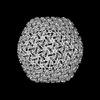
- Structure visualization
| Structure viewer | Molecule: MolmilJmol/JSmol |
|---|
- Downloads & links
-Download
| PDBx/mmCIF format | 8rqb.cif.gz | 913 KB | Display | PDBx/mmCIF format |
|---|---|---|---|---|
| PDB format | pdb8rqb.ent.gz | 774.1 KB | Display | PDB format |
| PDBx/mmJSON format | 8rqb.json.gz | Tree view | PDBx/mmJSON format | |
| Others |  Other downloads Other downloads |
-Validation report
| Arichive directory | https://data.pdbj.org/pub/pdb/validation_reports/rq/8rqbftp://data.pdbj.org/pub/pdb/validation_reports/rq/8rqb | HTTPS FTP |
|---|
-Related structure data
| Related structure data |  19436MC C: citing same article ( M: map data used to model this data |
|---|---|
| Similar structure data | |
| Experimental dataset #1 | Data reference: 10.6019/EMPIAR-11866 / Data set type: EMPIAR / Metadata reference: 10.6019/EMPIAR-11866 |


-Links
 PDBj
PDBj


- Assembly
| Deposited unit | 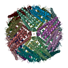
|
|---|---|
| 1 |
|
-Components
| #1: Protein | Mass: 20079.594 Da / Num. of mol.: 24 Source method: isolated from a genetically manipulated source Source: (gene. exp.)  #2: Chemical | ChemComp-FE /   Mass: 55.845 Da / Num. of mol.: 6 / Source method: obtained synthetically / Formula: Fe Mass: 55.845 Da / Num. of mol.: 6 / Source method: obtained synthetically / Formula: Fe#3: Chemical | ChemComp-ZN /   Mass: 65.409 Da / Num. of mol.: 24 / Source method: obtained synthetically / Formula: Zn Mass: 65.409 Da / Num. of mol.: 24 / Source method: obtained synthetically / Formula: Zn#4: Water | ChemComp-HOH / |  Mass: 18.015 Da / Num. of mol.: 2622 / Source method: isolated from a natural source / Formula: H2O Mass: 18.015 Da / Num. of mol.: 2622 / Source method: isolated from a natural source / Formula: H2OHas ligand of interest | N | |
|---|
-Experimental details
-Experiment
| Experiment | Method: ELECTRON MICROSCOPY |
|---|---|
| EM experiment | Aggregation state: PARTICLE / 3D reconstruction method: single particle reconstruction |
- Sample preparation
| Component | Name: Apoferritin from Mus musculus / Type: COMPLEX / Entity ID: #1 / Source: RECOMBINANT |
|---|---|
| Molecular weight | Experimental value: NO |
| Source (natural) | Organism: |
| Source (recombinant) | Organism: |
| Buffer solution | pH: 7.5 |
| Specimen | Conc.: 8 mg/ml / Embedding applied: NO / Shadowing applied: NO / Staining applied: NO / Vitrification applied: YES |
| Specimen support | Grid material: GOLD / Grid type: Quantifoil R1.2/1.3 |
| Vitrification | Instrument: LEICA EM GP / Cryogen name: ETHANE / Humidity: 100 % / Chamber temperature: 277 K |
- Electron microscopy imaging
| Experimental equipment |  Model: Titan Krios / Image courtesy: FEI Company |
|---|---|
| Microscopy | Model: FEI TITAN KRIOS |
| Electron gun | Electron source:  FIELD EMISSION GUN / Accelerating voltage: 300 kV / Illumination mode: FLOOD BEAM FIELD EMISSION GUN / Accelerating voltage: 300 kV / Illumination mode: FLOOD BEAM |
| Electron lens | Mode: BRIGHT FIELD / Nominal defocus max: 900 nm / Nominal defocus min: 300 nm |
| Specimen holder | Specimen holder model: FEI TITAN KRIOS AUTOGRID HOLDER |
| Image recording | Electron dose: 40 e/Å2 / Film or detector model: FEI FALCON IV (4k x 4k) |
- Processing
| EM software | Name: EPU / Version: 3 / Category: image acquisition |
|---|---|
| CTF correction | Type: PHASE FLIPPING AND AMPLITUDE CORRECTION |
| Symmetry | Point symmetry: O (octahedral) |
| 3D reconstruction | Resolution: 1.09 Å / Resolution method: FSC 0.143 CUT-OFF / Num. of particles: 411705 / Symmetry type: POINT |
