 Movie
Movie Controller


+ Open data

- Basic information
| Entry | Database: PDB / ID: 8rph | ||||||
|---|---|---|---|---|---|---|---|
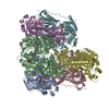
| Title | JanthE from Janthinobacterium sp. HH01,ketobutyryl-ThDP | ||||||
 Components Components | Thiamine pyrophosphate-binding protein | ||||||
 Keywords Keywords | TRANSFERASE / THDP / FAD | ||||||
| Function / homology | : / FLAVIN-ADENINE DINUCLEOTIDE / TRIETHYLENE GLYCOL Function and homology information Function and homology information | ||||||
| Biological species |  Janthinobacterium sp. HH01 (bacteria) Janthinobacterium sp. HH01 (bacteria) | ||||||
| Method |  X-RAY DIFFRACTION / SYNCHROTRON / MOLECULAR REPLACEMENT / Resolution: 2.96 Å X-RAY DIFFRACTION / SYNCHROTRON / MOLECULAR REPLACEMENT / Resolution: 2.96 Å | ||||||
 Authors Authors | Lanza, L. / Leogrande, C. / Rabe von Pappenheim, F. / Tittmann, K. / Mueller, M. | ||||||
| Funding support | European Union, 1items
| ||||||
 Citation Citation | Journal: Angew.Chem.Int.Ed.Engl. / Year: 2024 Title: Identification and Characterization of Thiamine Diphosphate-Dependent Lyases with an Unusual CDG Motif. Authors: Lanza, L. / Rabe von Pappenheim, F. / Bjarnesen, D. / Leogrande, C. / Paul, A. / Krug, L. / Tittmann, K. / Muller, M. | ||||||
| History |
|
- Structure visualization
| Structure viewer | Molecule: MolmilJmol/JSmol |
|---|
- Downloads & links
-Download
| PDBx/mmCIF format | 8rph.cif.gz | 2.1 MB | Display | PDBx/mmCIF format |
|---|---|---|---|---|
| PDB format | pdb8rph.ent.gz | Display | PDB format | |
| PDBx/mmJSON format | 8rph.json.gz | Tree view | PDBx/mmJSON format | |
| Others |  Other downloads Other downloads |
-Validation report
| Summary document | 8rph_validation.pdf.gz | 3.3 MB | Display | wwPDB validaton report |
|---|---|---|---|---|
| Full document | 8rph_full_validation.pdf.gz | 3.4 MB | Display | |
| Data in XML | 8rph_validation.xml.gz | 114.5 KB | Display | |
| Data in CIF | 8rph_validation.cif.gz | 151.9 KB | Display | |
| Arichive directory | https://data.pdbj.org/pub/pdb/validation_reports/rp/8rphftp://data.pdbj.org/pub/pdb/validation_reports/rp/8rph | HTTPS FTP |
-Related structure data

-Links
 PDBj
PDBj- Assembly
| Deposited unit | 
| ||||||||||||
|---|---|---|---|---|---|---|---|---|---|---|---|---|---|
| 1 | 
| ||||||||||||
| 2 | 
| ||||||||||||
| 3 | 
| ||||||||||||
| Unit cell |
|
-Components
-Protein , 1 types, 6 molecules ABCDEF
| #1: Protein | Mass: 68003.844 Da / Num. of mol.: 6 Source method: isolated from a genetically manipulated source Source: (gene. exp.) Janthinobacterium sp. HH01 (bacteria) / Production host: |
|---|
-Non-polymers , 5 types, 21 molecules 


| #2: Chemical | ChemComp-FAD /  Mass: 785.550 Da / Num. of mol.: 6 / Source method: obtained synthetically / Formula: C27H33N9O15P2 / Comment: FAD*YM Mass: 785.550 Da / Num. of mol.: 6 / Source method: obtained synthetically / Formula: C27H33N9O15P2 / Comment: FAD*YM#3: Chemical | ChemComp-MG /  Mass: 24.305 Da / Num. of mol.: 6 / Source method: obtained synthetically / Formula: Mg Mass: 24.305 Da / Num. of mol.: 6 / Source method: obtained synthetically / Formula: Mg#4: Chemical | ChemComp-A1H2A / ( Mass: 527.403 Da / Num. of mol.: 6 / Source method: obtained synthetically / Formula: C16H25N4O10P2S / Feature type: SUBJECT OF INVESTIGATION #5: Chemical | ChemComp-PGE / |  Mass: 150.173 Da / Num. of mol.: 1 / Source method: obtained synthetically / Formula: C6H14O4 Mass: 150.173 Da / Num. of mol.: 1 / Source method: obtained synthetically / Formula: C6H14O4#6: Water | ChemComp-HOH / | Mass: 18.015 Da / Num. of mol.: 2 / Source method: isolated from a natural source / Formula: H2O |
|---|
-Details
| Has ligand of interest | Y |
|---|
-Experimental details
-Experiment
| Experiment | Method: X-RAY DIFFRACTION / Number of used crystals: 1 |
|---|
- Sample preparation
| Crystal | Density Matthews: 2.55 Å3/Da / Density % sol: 51.78 % |
|---|---|
| Crystal grow | Temperature: 293 K / Method: vapor diffusion, hanging drop Details: Bis-Tris, Tris, Ammonium acetate, PEG 10000, Magnesium chloride, Thiamine diphosphate |
-Data collection
| Diffraction | Mean temperature: 100 K / Serial crystal experiment: N |
|---|---|
| Diffraction source | Source: SYNCHROTRON / Site: PETRA III, EMBL c/o DESY  / Beamline: P14 (MX2) / Wavelength: 0.68879 Å / Beamline: P14 (MX2) / Wavelength: 0.68879 Å |
| Detector | Type: DECTRIS EIGER2 X CdTe 16M / Detector: PIXEL / Date: Mar 13, 2023 |
| Radiation | Protocol: SINGLE WAVELENGTH / Monochromatic (M) / Laue (L): M / Scattering type: x-ray |
| Radiation wavelength | Wavelength: 0.68879 Å / Relative weight: 1 |
| Reflection | Resolution: 2.96→99.82 Å / Num. obs: 79335 / % possible obs: 94.5 % / Redundancy: 7 % / Biso Wilson estimate: 65.62 Å2 / CC1/2: 0.996 / Rrim(I) all: 0.144 / Net I/σ(I): 11.3 |
| Reflection shell | Resolution: 2.96→3.014 Å / Redundancy: 7.3 % / Mean I/σ(I) obs: 2.1 / Num. unique obs: 4130 / CC1/2: 0.701 / Rrim(I) all: 1.03 |
- Processing
| Software |
| |||||||||||||||||||||||||||||||||||||||||||||||||||||||||||||||||||||||||||||||||||||||||||||||||||||||||||||||||||||||||||||||||||||||||||||||||||||||||||||||||||||||||||||||||||||||||||||||||||||||||||
|---|---|---|---|---|---|---|---|---|---|---|---|---|---|---|---|---|---|---|---|---|---|---|---|---|---|---|---|---|---|---|---|---|---|---|---|---|---|---|---|---|---|---|---|---|---|---|---|---|---|---|---|---|---|---|---|---|---|---|---|---|---|---|---|---|---|---|---|---|---|---|---|---|---|---|---|---|---|---|---|---|---|---|---|---|---|---|---|---|---|---|---|---|---|---|---|---|---|---|---|---|---|---|---|---|---|---|---|---|---|---|---|---|---|---|---|---|---|---|---|---|---|---|---|---|---|---|---|---|---|---|---|---|---|---|---|---|---|---|---|---|---|---|---|---|---|---|---|---|---|---|---|---|---|---|---|---|---|---|---|---|---|---|---|---|---|---|---|---|---|---|---|---|---|---|---|---|---|---|---|---|---|---|---|---|---|---|---|---|---|---|---|---|---|---|---|---|---|---|---|---|---|---|---|---|
| Refinement | Method to determine structure: MOLECULAR REPLACEMENT / Resolution: 2.96→99.82 Å / SU ML: 0.3789 / Cross valid method: FREE R-VALUE / σ(F): 1.33 / Phase error: 25.628 Stereochemistry target values: GeoStd + Monomer Library + CDL v1.2
| |||||||||||||||||||||||||||||||||||||||||||||||||||||||||||||||||||||||||||||||||||||||||||||||||||||||||||||||||||||||||||||||||||||||||||||||||||||||||||||||||||||||||||||||||||||||||||||||||||||||||||
| Solvent computation | Shrinkage radii: 0.8 Å / VDW probe radii: 1 Å / Solvent model: FLAT BULK SOLVENT MODEL | |||||||||||||||||||||||||||||||||||||||||||||||||||||||||||||||||||||||||||||||||||||||||||||||||||||||||||||||||||||||||||||||||||||||||||||||||||||||||||||||||||||||||||||||||||||||||||||||||||||||||||
| Displacement parameters | Biso mean: 71.61 Å2 | |||||||||||||||||||||||||||||||||||||||||||||||||||||||||||||||||||||||||||||||||||||||||||||||||||||||||||||||||||||||||||||||||||||||||||||||||||||||||||||||||||||||||||||||||||||||||||||||||||||||||||
| Refinement step | Cycle: LAST / Resolution: 2.96→99.82 Å
| |||||||||||||||||||||||||||||||||||||||||||||||||||||||||||||||||||||||||||||||||||||||||||||||||||||||||||||||||||||||||||||||||||||||||||||||||||||||||||||||||||||||||||||||||||||||||||||||||||||||||||
| Refine LS restraints |
| |||||||||||||||||||||||||||||||||||||||||||||||||||||||||||||||||||||||||||||||||||||||||||||||||||||||||||||||||||||||||||||||||||||||||||||||||||||||||||||||||||||||||||||||||||||||||||||||||||||||||||
| LS refinement shell |
| |||||||||||||||||||||||||||||||||||||||||||||||||||||||||||||||||||||||||||||||||||||||||||||||||||||||||||||||||||||||||||||||||||||||||||||||||||||||||||||||||||||||||||||||||||||||||||||||||||||||||||
| Refinement TLS params. | Method: refined / Origin x: 40.4727206368 Å / Origin y: -14.2401914575 Å / Origin z: 49.8534235079 Å
| |||||||||||||||||||||||||||||||||||||||||||||||||||||||||||||||||||||||||||||||||||||||||||||||||||||||||||||||||||||||||||||||||||||||||||||||||||||||||||||||||||||||||||||||||||||||||||||||||||||||||||
| Refinement TLS group | Selection details: all |
