 Movie
Movie Controller


+ Open data

- Basic information
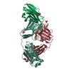
| Entry | Database: PDB / ID: 8rp8 | ||||||
|---|---|---|---|---|---|---|---|
| Title | Structure of K2 Fab in complex with human CD47 ECD | ||||||
 Components Components |
| ||||||
 Keywords Keywords | IMMUNE SYSTEM / Fab / glycoprotein | ||||||
| Function / homology |  Function and homology information Function and homology informationcellular response to interleukin-12 / regulation of Fc receptor mediated stimulatory signaling pathway / protein binding involved in heterotypic cell-cell adhesion / positive regulation of monocyte extravasation / regulation of type II interferon production / ATP export / positive regulation of cell-cell adhesion / regulation of interleukin-10 production / cell-cell adhesion mediator activity / regulation of tumor necrosis factor production ...cellular response to interleukin-12 / regulation of Fc receptor mediated stimulatory signaling pathway / protein binding involved in heterotypic cell-cell adhesion / positive regulation of monocyte extravasation / regulation of type II interferon production / ATP export / positive regulation of cell-cell adhesion / regulation of interleukin-10 production / cell-cell adhesion mediator activity / regulation of tumor necrosis factor production / regulation of interleukin-12 production / regulation of nitric oxide biosynthetic process / negative regulation of phagocytosis / regulation of interleukin-6 production / Signal regulatory protein family interactions / thrombospondin receptor activity / tertiary granule membrane / cellular response to interleukin-1 / Integrin cell surface interactions / specific granule membrane / positive regulation of phagocytosis / positive regulation of stress fiber assembly / integrin-mediated signaling pathway / Cell surface interactions at the vascular wall / cellular response to type II interferon / positive regulation of inflammatory response / cell migration / positive regulation of T cell activation / angiogenesis / inflammatory response / apoptotic process / positive regulation of cell population proliferation / Neutrophil degranulation / cell surface / extracellular exosome / plasma membrane Similarity search - Function | ||||||
| Biological species |  Homo sapiens (human) Homo sapiens (human) | ||||||
| Method |  X-RAY DIFFRACTION / SYNCHROTRON / MOLECULAR REPLACEMENT / Resolution: 2 Å X-RAY DIFFRACTION / SYNCHROTRON / MOLECULAR REPLACEMENT / Resolution: 2 Å | ||||||
 Authors Authors | Laursen, M. / Kelpsas, V. / Rose, N. | ||||||
| Funding support |  Switzerland, 1items Switzerland, 1items
| ||||||
 Citation Citation | Journal: Mabs / Year: 2024 Title: Structural analysis of light chain-driven bispecific antibodies targeting CD47 and PD-L1. Authors: Malinge, P. / Chauchet, X. / Bourguignon, J. / Bosson, N. / Calloud, S. / Bautzova, T. / Borlet, M. / Laursen, M. / Kelpsas, V. / Rose, N. / Gueneau, F. / Ravn, U. / Magistrelli, G. / Fischer, N. | ||||||
| History |
|
- Structure visualization
| Structure viewer | Molecule: MolmilJmol/JSmol |
|---|
- Downloads & links
-Download
| PDBx/mmCIF format | 8rp8.cif.gz | 442.8 KB | Display | PDBx/mmCIF format |
|---|---|---|---|---|
| PDB format | pdb8rp8.ent.gz | 363.8 KB | Display | PDB format |
| PDBx/mmJSON format | 8rp8.json.gz | Tree view | PDBx/mmJSON format | |
| Others |  Other downloads Other downloads |
-Validation report
| Summary document | 8rp8_validation.pdf.gz | 512.7 KB | Display | wwPDB validaton report |
|---|---|---|---|---|
| Full document | 8rp8_full_validation.pdf.gz | 519.2 KB | Display | |
| Data in XML | 8rp8_validation.xml.gz | 48.2 KB | Display | |
| Data in CIF | 8rp8_validation.cif.gz | 70.1 KB | Display | |
| Arichive directory | https://data.pdbj.org/pub/pdb/validation_reports/rp/8rp8ftp://data.pdbj.org/pub/pdb/validation_reports/rp/8rp8 | HTTPS FTP |
-Related structure data
-Links
 PDBj
PDBj


- Assembly
| Deposited unit | 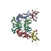
| ||||||||
|---|---|---|---|---|---|---|---|---|---|
| 1 |
| ||||||||
| 2 |
| ||||||||
| Unit cell |
|
-Components
-Antibody , 2 types, 4 molecules AHBL
| #1: Antibody | Mass: 23468.174 Da / Num. of mol.: 2 Source method: isolated from a genetically manipulated source Source: (gene. exp.) Homo sapiens (human) / Production host: Homo sapiens (human)#2: Antibody | Mass: 23472.129 Da / Num. of mol.: 2 Source method: isolated from a genetically manipulated source Source: (gene. exp.) Homo sapiens (human) / Production host: Homo sapiens (human) |
|---|
-Protein / Sugars , 2 types, 8 molecules CD
| #3: Protein | Mass: 14747.510 Da / Num. of mol.: 2 Source method: isolated from a genetically manipulated source Source: (gene. exp.) Homo sapiens (human) / Gene: CD47, MER6 / Production host: Homo sapiens (human) / References: UniProt: Q08722#6: Sugar | ChemComp-NAG /  Type: D-saccharide, beta linking / Mass: 221.208 Da / Num. of mol.: 6 / Source method: isolated from a natural source / Formula: C8H15NO6 Type: D-saccharide, beta linking / Mass: 221.208 Da / Num. of mol.: 6 / Source method: isolated from a natural source / Formula: C8H15NO6 |
|---|
-Non-polymers , 3 types, 822 molecules 


| #4: Chemical |  Mass: 106.120 Da / Num. of mol.: 2 / Source method: obtained synthetically / Formula: C4H10O3 Mass: 106.120 Da / Num. of mol.: 2 / Source method: obtained synthetically / Formula: C4H10O3#5: Chemical |  Mass: 92.094 Da / Num. of mol.: 2 / Source method: isolated from a natural source / Formula: C3H8O3 Mass: 92.094 Da / Num. of mol.: 2 / Source method: isolated from a natural source / Formula: C3H8O3#7: Water | ChemComp-HOH / | Mass: 18.015 Da / Num. of mol.: 818 / Source method: isolated from a natural source / Formula: H2O |
|---|
-Details
| Has ligand of interest | N |
|---|
-Experimental details
-Experiment
| Experiment | Method: X-RAY DIFFRACTION / Number of used crystals: 1 |
|---|
- Sample preparation
| Crystal | Density Matthews: 2.61 Å3/Da / Density % sol: 52.9 % |
|---|---|
| Crystal grow | Temperature: 293 K / Method: vapor diffusion, sitting drop / pH: 5 / Details: 0.1 M sodium citrate, 20% PEG 8000 |
-Data collection
| Diffraction | Mean temperature: 100 K / Serial crystal experiment: N |
|---|---|
| Diffraction source | Source: SYNCHROTRON / Site: MAX IV  / Beamline: BioMAX / Wavelength: 0.9763 Å / Beamline: BioMAX / Wavelength: 0.9763 Å |
| Detector | Type: DECTRIS EIGER X 16M / Detector: PIXEL / Date: Nov 18, 2021 |
| Radiation | Monochromator: Si(111) HDCM / Protocol: SINGLE WAVELENGTH / Monochromatic (M) / Laue (L): M / Scattering type: x-ray |
| Radiation wavelength | Wavelength: 0.9763 Å / Relative weight: 1 |
| Reflection | Resolution: 2→70.22 Å / Num. obs: 78163 / % possible obs: 97.4 % / Redundancy: 3.6 % / CC1/2: 0.999 / Rmerge(I) obs: 0.036 / Rpim(I) all: 0.022 / Rrim(I) all: 0.043 / Χ2: 0.97 / Net I/σ(I): 19.2 |
| Reflection shell | Resolution: 2→2.04 Å / Redundancy: 3.7 % / Rmerge(I) obs: 0.145 / Mean I/σ(I) obs: 7.5 / Num. unique obs: 4427 / CC1/2: 0.981 / Rpim(I) all: 0.089 / Rrim(I) all: 0.171 / Χ2: 1.07 / % possible all: 96.8 |
- Processing
| Software |
| |||||||||||||||||||||||||||||||||||||||||||||||||||||||||||||||||||||||||||||||||||||||||||||||||||||||||||||||||||||||||||||||||||||||||||||||||||||||||||||||||||||||||||||||
|---|---|---|---|---|---|---|---|---|---|---|---|---|---|---|---|---|---|---|---|---|---|---|---|---|---|---|---|---|---|---|---|---|---|---|---|---|---|---|---|---|---|---|---|---|---|---|---|---|---|---|---|---|---|---|---|---|---|---|---|---|---|---|---|---|---|---|---|---|---|---|---|---|---|---|---|---|---|---|---|---|---|---|---|---|---|---|---|---|---|---|---|---|---|---|---|---|---|---|---|---|---|---|---|---|---|---|---|---|---|---|---|---|---|---|---|---|---|---|---|---|---|---|---|---|---|---|---|---|---|---|---|---|---|---|---|---|---|---|---|---|---|---|---|---|---|---|---|---|---|---|---|---|---|---|---|---|---|---|---|---|---|---|---|---|---|---|---|---|---|---|---|---|---|---|---|---|
| Refinement | Method to determine structure: MOLECULAR REPLACEMENT / Resolution: 2→20.06 Å / Cor.coef. Fo:Fc: 0.951 / Cor.coef. Fo:Fc free: 0.932 / SU R Cruickshank DPI: 0.173 / Cross valid method: THROUGHOUT / SU R Blow DPI: 0.177 / SU Rfree Blow DPI: 0.149 / SU Rfree Cruickshank DPI: 0.149
| |||||||||||||||||||||||||||||||||||||||||||||||||||||||||||||||||||||||||||||||||||||||||||||||||||||||||||||||||||||||||||||||||||||||||||||||||||||||||||||||||||||||||||||||
| Displacement parameters | Biso mean: 37.02 Å2
| |||||||||||||||||||||||||||||||||||||||||||||||||||||||||||||||||||||||||||||||||||||||||||||||||||||||||||||||||||||||||||||||||||||||||||||||||||||||||||||||||||||||||||||||
| Refine analyze | Luzzati coordinate error obs: 0.23 Å | |||||||||||||||||||||||||||||||||||||||||||||||||||||||||||||||||||||||||||||||||||||||||||||||||||||||||||||||||||||||||||||||||||||||||||||||||||||||||||||||||||||||||||||||
| Refinement step | Cycle: LAST / Resolution: 2→20.06 Å
| |||||||||||||||||||||||||||||||||||||||||||||||||||||||||||||||||||||||||||||||||||||||||||||||||||||||||||||||||||||||||||||||||||||||||||||||||||||||||||||||||||||||||||||||
| Refine LS restraints |
| |||||||||||||||||||||||||||||||||||||||||||||||||||||||||||||||||||||||||||||||||||||||||||||||||||||||||||||||||||||||||||||||||||||||||||||||||||||||||||||||||||||||||||||||
| LS refinement shell | Resolution: 2→2.01 Å
| |||||||||||||||||||||||||||||||||||||||||||||||||||||||||||||||||||||||||||||||||||||||||||||||||||||||||||||||||||||||||||||||||||||||||||||||||||||||||||||||||||||||||||||||
| Refinement TLS params. | Refine-ID: X-RAY DIFFRACTION
| |||||||||||||||||||||||||||||||||||||||||||||||||||||||||||||||||||||||||||||||||||||||||||||||||||||||||||||||||||||||||||||||||||||||||||||||||||||||||||||||||||||||||||||||
| Refinement TLS group |
|
