Journal: Protein Sci / Year: 2024 Title: Molecular basis for different substrate-binding sites and chaperone functions of the BRICHOS domain. Authors: Gefei Chen / Yu Wang / Zihan Zheng / Wangshu Jiang / Axel Leppert / Xueying Zhong / Anna Belorusova / Gregg Siegal / Caroline Jegerschöld / Philip J B Koeck / Axel Abelein / Hans Hebert / ...Authors: Gefei Chen / Yu Wang / Zihan Zheng / Wangshu Jiang / Axel Leppert / Xueying Zhong / Anna Belorusova / Gregg Siegal / Caroline Jegerschöld / Philip J B Koeck / Axel Abelein / Hans Hebert / Stefan D Knight / Jan Johansson / Abstract: Proteins can misfold into fibrillar or amorphous aggregates and molecular chaperones act as crucial guardians against these undesirable processes. The BRICHOS chaperone domain, found in several ...Proteins can misfold into fibrillar or amorphous aggregates and molecular chaperones act as crucial guardians against these undesirable processes. The BRICHOS chaperone domain, found in several otherwise unrelated proproteins that contain amyloidogenic regions, effectively inhibits amyloid formation and toxicity but can in some cases also prevent non-fibrillar, amorphous protein aggregation. Here, we elucidate the molecular basis behind the multifaceted chaperone activities of the BRICHOS domain from the Bri2 proprotein. High-confidence AlphaFold2 and RoseTTAFold predictions suggest that the intramolecular amyloidogenic region (Bri23) is part of the hydrophobic core of the proprotein, where it occupies the proposed amyloid binding site, explaining the markedly reduced ability of the proprotein to prevent an exogenous amyloidogenic peptide from aggregating. However, the BRICHOS-Bri23 complex maintains its ability to form large polydisperse oligomers that prevent amorphous protein aggregation. A cryo-EM-derived model of the Bri2 BRICHOS oligomer is compatible with surface-exposed hydrophobic motifs that get exposed and come together during oligomerization, explaining its effects against amorphous aggregation. These findings provide a molecular basis for the BRICHOS chaperone domain function, where distinct surfaces are employed against different forms of protein aggregation.
A: Integral membrane protein 2B K: Integral membrane protein 2B E: Integral membrane protein 2B I: Integral membrane protein 2B C: Integral membrane protein 2B G: Integral membrane protein 2B B: Integral membrane protein 2B H: Integral membrane protein 2B D: Integral membrane protein 2B J: Integral membrane protein 2B F: Integral membrane protein 2B L: Integral membrane protein 2B
In the structure databanks used in Yorodumi, some data are registered as the other names, "COVID-19 virus" and "2019-nCoV". Here are the details of the virus and the list of structure data.
Jan 31, 2019. EMDB accession codes are about to change! (news from PDBe EMDB page)
EMDB accession codes are about to change! (news from PDBe EMDB page)
The allocation of 4 digits for EMDB accession codes will soon come to an end. Whilst these codes will remain in use, new EMDB accession codes will include an additional digit and will expand incrementally as the available range of codes is exhausted. The current 4-digit format prefixed with “EMD-” (i.e. EMD-XXXX) will advance to a 5-digit format (i.e. EMD-XXXXX), and so on. It is currently estimated that the 4-digit codes will be depleted around Spring 2019, at which point the 5-digit format will come into force.
The EM Navigator/Yorodumi systems omit the EMD- prefix.
Related info.:Q: What is EMD? / ID/Accession-code notation in Yorodumi/EM Navigator
Yorodumi is a browser for structure data from EMDB, PDB, SASBDB, etc.
This page is also the successor to EM Navigator detail page, and also detail information page/front-end page for Omokage search.
The word "yorodu" (or yorozu) is an old Japanese word meaning "ten thousand". "mi" (miru) is to see.
Related info.:EMDB / PDB / SASBDB / Comparison of 3 databanks / Yorodumi Search / Aug 31, 2016. New EM Navigator & Yorodumi / Yorodumi Papers / Jmol/JSmol / Function and homology information / Changes in new EM Navigator and Yorodumi
 Movie
Movie Controller


 Open data
 Basic information
 Components
 Keywords
 Function and homology information
 Homo sapiens (human)
 Authors
 Sweden, 1items
  Citation

 Structure visualization
 Downloads & links
 Other downloads
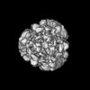
 PDBj
PDBj
 Assembly
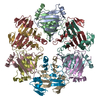

 Sample preparation
 Electron microscopy imaging
 FIELD EMISSION GUN / Accelerating voltage: 300 kV / Illumination mode: FLOOD BEAM
 Processing