 Movie
Movie Controller


+ Open data

- Basic information
| Entry | Database: PDB / ID: 8rly | ||||||
|---|---|---|---|---|---|---|---|
| Title | E. coli endonuclease IV complexed with sulfate, catalytic Fe2+ | ||||||
 Components Components | Endonuclease 4 | ||||||
 Keywords Keywords | DNA BINDING PROTEIN / bacterial endonuclease IV / catalytic iron | ||||||
| Function / homology |  Function and homology information Function and homology informationdeoxyribonuclease IV / deoxyribonuclease IV (phage-T4-induced) activity / 3'-5'-DNA exonuclease activity / phosphoric diester hydrolase activity / phosphatase activity / DNA-(apurinic or apyrimidinic site) endonuclease activity / base-excision repair / endonuclease activity / DNA repair / DNA binding ...deoxyribonuclease IV / deoxyribonuclease IV (phage-T4-induced) activity / 3'-5'-DNA exonuclease activity / phosphoric diester hydrolase activity / phosphatase activity / DNA-(apurinic or apyrimidinic site) endonuclease activity / base-excision repair / endonuclease activity / DNA repair / DNA binding / zinc ion binding / cytosol Similarity search - Function | ||||||
| Biological species |  | ||||||
| Method |  X-RAY DIFFRACTION / SYNCHROTRON / MOLECULAR REPLACEMENT / Resolution: 1.9 Å X-RAY DIFFRACTION / SYNCHROTRON / MOLECULAR REPLACEMENT / Resolution: 1.9 Å | ||||||
 Authors Authors | Saper, M.A. / Paterson, N.G. / Kirillov, S. / Rouvinski, A. | ||||||
| Funding support | 1items
| ||||||
 Citation Citation | Journal: Biochemistry / Year: 2025 Title: Octahedral Iron in Catalytic Sites of Endonuclease IV from Staphylococcus aureus and Escherichia coli . Authors: Kirillov, S. / Isupov, M. / Paterson, N.G. / Wiener, R. / Abeldenov, S. / Saper, M.A. / Rouvinski, A. #1: Journal: Acta Crystallogr D Struct Biol / Year: 2019 Title: Macromolecular structure determination using X-rays, neutrons and electrons: recent developments in Phenix. Authors: Dorothee Liebschner / Pavel V Afonine / Matthew L Baker / Gábor Bunkóczi / Vincent B Chen / Tristan I Croll / Bradley Hintze / Li Wei Hung / Swati Jain / Airlie J McCoy / Nigel W Moriarty ...Authors: Dorothee Liebschner / Pavel V Afonine / Matthew L Baker / Gábor Bunkóczi / Vincent B Chen / Tristan I Croll / Bradley Hintze / Li Wei Hung / Swati Jain / Airlie J McCoy / Nigel W Moriarty / Robert D Oeffner / Billy K Poon / Michael G Prisant / Randy J Read / Jane S Richardson / David C Richardson / Massimo D Sammito / Oleg V Sobolev / Duncan H Stockwell / Thomas C Terwilliger / Alexandre G Urzhumtsev / Lizbeth L Videau / Christopher J Williams / Paul D Adams /    Abstract: Diffraction (X-ray, neutron and electron) and electron cryo-microscopy are powerful methods to determine three-dimensional macromolecular structures, which are required to understand biological ...Diffraction (X-ray, neutron and electron) and electron cryo-microscopy are powerful methods to determine three-dimensional macromolecular structures, which are required to understand biological processes and to develop new therapeutics against diseases. The overall structure-solution workflow is similar for these techniques, but nuances exist because the properties of the reduced experimental data are different. Software tools for structure determination should therefore be tailored for each method. Phenix is a comprehensive software package for macromolecular structure determination that handles data from any of these techniques. Tasks performed with Phenix include data-quality assessment, map improvement, model building, the validation/rebuilding/refinement cycle and deposition. Each tool caters to the type of experimental data. The design of Phenix emphasizes the automation of procedures, where possible, to minimize repetitive and time-consuming manual tasks, while default parameters are chosen to encourage best practice. A graphical user interface provides access to many command-line features of Phenix and streamlines the transition between programs, project tracking and re-running of previous tasks. | ||||||
| History |
|
- Structure visualization
| Structure viewer | Molecule: MolmilJmol/JSmol |
|---|
- Downloads & links
-Download
| PDBx/mmCIF format | 8rly.cif.gz | 664.3 KB | Display | PDBx/mmCIF format |
|---|---|---|---|---|
| PDB format | pdb8rly.ent.gz | 442.4 KB | Display | PDB format |
| PDBx/mmJSON format | 8rly.json.gz | Tree view | PDBx/mmJSON format | |
| Others |  Other downloads Other downloads |
-Validation report
| Summary document | 8rly_validation.pdf.gz | 11.9 MB | Display | wwPDB validaton report |
|---|---|---|---|---|
| Full document | 8rly_full_validation.pdf.gz | 11.8 MB | Display | |
| Data in XML | 8rly_validation.xml.gz | 69.8 KB | Display | |
| Data in CIF | 8rly_validation.cif.gz | 97 KB | Display | |
| Arichive directory | https://data.pdbj.org/pub/pdb/validation_reports/rl/8rlyftp://data.pdbj.org/pub/pdb/validation_reports/rl/8rly | HTTPS FTP |
-Related structure data
| Related structure data | 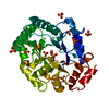 8axyC 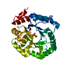 8eddC 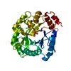 8pkbC 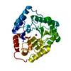 1qtwS C: citing same article ( S: Starting model for refinement |
|---|---|
| Similar structure data |
-Links
 PDBj
PDBj


- Assembly
| Deposited unit | 
| ||||||||||||
|---|---|---|---|---|---|---|---|---|---|---|---|---|---|
| 1 | 
| ||||||||||||
| 2 | 
| ||||||||||||
| 3 | 
| ||||||||||||
| 4 | 
| ||||||||||||
| 5 | 
| ||||||||||||
| Unit cell |
|
-Components
-Protein , 1 types, 5 molecules ABCDE
| #1: Protein | Mass: 31518.496 Da / Num. of mol.: 5 Source method: isolated from a genetically manipulated source Details: C-terminus not resolved in some of the chains. / Source: (gene. exp.) |
|---|
-Non-polymers , 7 types, 1151 molecules 


| #2: Chemical | ChemComp-ZN /  Mass: 65.409 Da / Num. of mol.: 10 / Source method: obtained synthetically / Formula: Zn / Feature type: SUBJECT OF INVESTIGATION Mass: 65.409 Da / Num. of mol.: 10 / Source method: obtained synthetically / Formula: Zn / Feature type: SUBJECT OF INVESTIGATION#3: Chemical | ChemComp-FE2 /  Mass: 55.845 Da / Num. of mol.: 5 / Source method: obtained synthetically / Formula: Fe / Feature type: SUBJECT OF INVESTIGATION Mass: 55.845 Da / Num. of mol.: 5 / Source method: obtained synthetically / Formula: Fe / Feature type: SUBJECT OF INVESTIGATION#4: Chemical | ChemComp-EPE / |  Mass: 238.305 Da / Num. of mol.: 1 / Source method: obtained synthetically / Formula: C8H18N2O4S / Comment: pH buffer*YM Mass: 238.305 Da / Num. of mol.: 1 / Source method: obtained synthetically / Formula: C8H18N2O4S / Comment: pH buffer*YM#5: Chemical | ChemComp-SO4 /  Mass: 96.063 Da / Num. of mol.: 5 / Source method: obtained synthetically / Formula: SO4 / Feature type: SUBJECT OF INVESTIGATION Mass: 96.063 Da / Num. of mol.: 5 / Source method: obtained synthetically / Formula: SO4 / Feature type: SUBJECT OF INVESTIGATION#6: Chemical | ChemComp-NI /  Mass: 58.693 Da / Num. of mol.: 7 / Source method: obtained synthetically / Formula: Ni / Feature type: SUBJECT OF INVESTIGATION Mass: 58.693 Da / Num. of mol.: 7 / Source method: obtained synthetically / Formula: Ni / Feature type: SUBJECT OF INVESTIGATION#7: Chemical | ChemComp-CL /  Mass: 35.453 Da / Num. of mol.: 5 / Source method: isolated from a natural source / Formula: Cl Mass: 35.453 Da / Num. of mol.: 5 / Source method: isolated from a natural source / Formula: Cl#8: Water | ChemComp-HOH / | Mass: 18.015 Da / Num. of mol.: 1118 / Source method: isolated from a natural source / Formula: H2O |
|---|
-Details
| Has ligand of interest | Y |
|---|---|
| Has protein modification | N |
-Experimental details
-Experiment
| Experiment | Method: X-RAY DIFFRACTION / Number of used crystals: 1 |
|---|
- Sample preparation
| Crystal | Density Matthews: 2.28 Å3/Da / Density % sol: 46.09 % |
|---|---|
| Crystal grow | Temperature: 295 K / Method: vapor diffusion, hanging drop Details: 1 microliter protein mixed with 2 microliter precipitant. Protein: 12.5 mg/ml, 20 mM Tris-HCl pH 8.0, 150 mM NaCl. Precipitant: 0.1 M HEPES pH 7.0, 15% Polyethylene Glycol 3,350, 10 mM ...Details: 1 microliter protein mixed with 2 microliter precipitant. Protein: 12.5 mg/ml, 20 mM Tris-HCl pH 8.0, 150 mM NaCl. Precipitant: 0.1 M HEPES pH 7.0, 15% Polyethylene Glycol 3,350, 10 mM Magnesium chloride hexahydrate, 5 mM nickel (II) chloride hexahydrate |
-Data collection
| Diffraction |
| |||||||||||||||||||||||||||||||||||
|---|---|---|---|---|---|---|---|---|---|---|---|---|---|---|---|---|---|---|---|---|---|---|---|---|---|---|---|---|---|---|---|---|---|---|---|---|
| Diffraction source |
| |||||||||||||||||||||||||||||||||||
| Detector |
| |||||||||||||||||||||||||||||||||||
| Radiation |
| |||||||||||||||||||||||||||||||||||
| Radiation wavelength |
| |||||||||||||||||||||||||||||||||||
| Reflection | Resolution: 1.9→97.12 Å / Num. obs: 193287 / % possible obs: 87.8 % / Redundancy: 5.8 % / Biso Wilson estimate: 22.18 Å2 / CC1/2: 0.998 / Rmerge(I) obs: 0.06475 / Rrim(I) all: 0.07067 / Net I/av σ(I): 14.86 / Net I/σ(I): 14.86 | |||||||||||||||||||||||||||||||||||
| Reflection shell | Resolution: 1.9→1.92 Å / Redundancy: 1.4 % / Rmerge(I) obs: 0.3256 / Mean I/σ(I) obs: 1.36 / Num. unique obs: 372 / CC1/2: 0.772 / Rpim(I) all: 0.3083 / Rrim(I) all: 0.4496 / % possible all: 21.7 |
- Processing
| Software |
| |||||||||||||||||||||||||||||||||||||||||||||||||||||||||||||||||||||||||||||||||||||||||||||||||||||||||||||||||||||||||||||||||||||||||||||||||||||||||||||||||||||||||||||||||||||||||||||||||||||||||||||||||||||||||
|---|---|---|---|---|---|---|---|---|---|---|---|---|---|---|---|---|---|---|---|---|---|---|---|---|---|---|---|---|---|---|---|---|---|---|---|---|---|---|---|---|---|---|---|---|---|---|---|---|---|---|---|---|---|---|---|---|---|---|---|---|---|---|---|---|---|---|---|---|---|---|---|---|---|---|---|---|---|---|---|---|---|---|---|---|---|---|---|---|---|---|---|---|---|---|---|---|---|---|---|---|---|---|---|---|---|---|---|---|---|---|---|---|---|---|---|---|---|---|---|---|---|---|---|---|---|---|---|---|---|---|---|---|---|---|---|---|---|---|---|---|---|---|---|---|---|---|---|---|---|---|---|---|---|---|---|---|---|---|---|---|---|---|---|---|---|---|---|---|---|---|---|---|---|---|---|---|---|---|---|---|---|---|---|---|---|---|---|---|---|---|---|---|---|---|---|---|---|---|---|---|---|---|---|---|---|---|---|---|---|---|---|---|---|---|---|---|---|---|
| Refinement | Method to determine structure: MOLECULAR REPLACEMENT Starting model: 1QTW Resolution: 1.9→97.12 Å / SU ML: 0.1741 / Cross valid method: FREE R-VALUE / σ(F): 1.35 / Phase error: 21.4556 Stereochemistry target values: GeoStd + Monomer Library + CDL v1.2
| |||||||||||||||||||||||||||||||||||||||||||||||||||||||||||||||||||||||||||||||||||||||||||||||||||||||||||||||||||||||||||||||||||||||||||||||||||||||||||||||||||||||||||||||||||||||||||||||||||||||||||||||||||||||||
| Solvent computation | Shrinkage radii: 0.9 Å / VDW probe radii: 1.1 Å / Solvent model: FLAT BULK SOLVENT MODEL | |||||||||||||||||||||||||||||||||||||||||||||||||||||||||||||||||||||||||||||||||||||||||||||||||||||||||||||||||||||||||||||||||||||||||||||||||||||||||||||||||||||||||||||||||||||||||||||||||||||||||||||||||||||||||
| Displacement parameters | Biso mean: 23.66 Å2 | |||||||||||||||||||||||||||||||||||||||||||||||||||||||||||||||||||||||||||||||||||||||||||||||||||||||||||||||||||||||||||||||||||||||||||||||||||||||||||||||||||||||||||||||||||||||||||||||||||||||||||||||||||||||||
| Refinement step | Cycle: LAST / Resolution: 1.9→97.12 Å
| |||||||||||||||||||||||||||||||||||||||||||||||||||||||||||||||||||||||||||||||||||||||||||||||||||||||||||||||||||||||||||||||||||||||||||||||||||||||||||||||||||||||||||||||||||||||||||||||||||||||||||||||||||||||||
| Refine LS restraints |
| |||||||||||||||||||||||||||||||||||||||||||||||||||||||||||||||||||||||||||||||||||||||||||||||||||||||||||||||||||||||||||||||||||||||||||||||||||||||||||||||||||||||||||||||||||||||||||||||||||||||||||||||||||||||||
| LS refinement shell |
|
