- PDB-8axy: Staphylococcus aureus endonuclease IV orthorhombic crystal form -
+
データを開く
IDまたはキーワード:
読み込み中...
-
基本情報
登録情報
データベース: PDB / ID: 8axy
タイトル
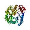
Staphylococcus aureus endonuclease IV orthorhombic crystal form
要素
Probable endonuclease 4
キーワード
HYDROLASE / endonuclease 4 / DNA repair / metalloenzyme
機能・相同性
機能・相同性情報
deoxyribonuclease IV / deoxyribonuclease IV (phage-T4-induced) activity / phosphoric diester hydrolase activity / DNA-(apurinic or apyrimidinic site) endonuclease activity / base-excision repair / DNA binding / zinc ion binding 類似検索 - 分子機能
AP endonucleases family 2 signature 1. / AP endonucleases family 2 signature 2. / AP endonuclease 2, zinc binding site / AP endonucleases family 2 signature 3. / AP endonucleases family 2 profile. / AP endonuclease family 2 / AP endonuclease 2 / Divalent-metal-dependent TIM barrel enzymes / Xylose isomerase-like, TIM barrel domain / Xylose isomerase-like TIM barrel ...AP endonucleases family 2 signature 1. / AP endonucleases family 2 signature 2. / AP endonuclease 2, zinc binding site / AP endonucleases family 2 signature 3. / AP endonucleases family 2 profile. / AP endonuclease family 2 / AP endonuclease 2 / Divalent-metal-dependent TIM barrel enzymes / Xylose isomerase-like, TIM barrel domain / Xylose isomerase-like TIM barrel / Xylose isomerase-like superfamily / TIM Barrel / Alpha-Beta Barrel / Alpha Beta 類似検索 - ドメイン・相同性
 ムービー
ムービー コントローラー


 データを開く
 基本情報
基本情報 要素
要素 キーワード
キーワード 機能・相同性情報
 Staphylococcus aureus (黄色ブドウ球菌)
 X線回折 /
  データ登録者
 イスラエル, Kazakhstan, 2件
  引用
引用 構造の表示
構造の表示 ダウンロードとリンク
 その他のダウンロード
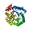


 PDBj
PDBj
 集合体
集合体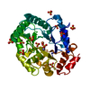

 分子量: 55.845 Da / 分子数: 2 / 由来タイプ: 合成 / 式: Fe / タイプ: SUBJECT OF INVESTIGATION
 分子量: 65.409 Da / 分子数: 1 / 由来タイプ: 合成 / 式: Zn / タイプ: SUBJECT OF INVESTIGATION
 分子量: 96.063 Da / 分子数: 12 / 由来タイプ: 合成 / 式: SO4 / タイプ: SUBJECT OF INVESTIGATION
 分子量: 18.015 Da / 分子数: 521 / 由来タイプ: 天然 / 式: H2O
 試料調製
試料調製 / ビームライン: I04 / 波長: 0.8 Å
 解析
解析