 Movie
Movie Controller


+ Open data

- Basic information
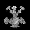
| Entry | Database: PDB / ID: 8rk8 | |||||||||
|---|---|---|---|---|---|---|---|---|---|---|
| Title | Tail of bacteriophage JBD30 computed in C6 symmetry | |||||||||
 Components Components | Major tail protein | |||||||||
 Keywords Keywords | VIRUS / bacteriophage JBD30 / virion / tail / major tail protein | |||||||||
| Function / homology | Protein of unknown function UCP028589 / Virion structural protein Function and homology information Function and homology information | |||||||||
| Biological species |  Pseudomonas phage JBD30 (virus) Pseudomonas phage JBD30 (virus) | |||||||||
| Method | ELECTRON MICROSCOPY / single particle reconstruction / cryo EM / Resolution: 3.88 Å | |||||||||
 Authors Authors | Valentova, L. / Fuzik, T. / Plevka, P. | |||||||||
| Funding support |  Czech Republic, European Union, 2items Czech Republic, European Union, 2items
| |||||||||
 Citation Citation | Journal: EMBO J / Year: 2024 Title: Structure and replication of Pseudomonas aeruginosa phage JBD30. Authors: Lucie Valentová / Tibor Füzik / Jiří Nováček / Zuzana Hlavenková / Jakub Pospíšil / Pavel Plevka / Abstract: Bacteriophages are the most abundant biological entities on Earth, but our understanding of many aspects of their lifecycles is still incomplete. Here, we have structurally analysed the infection ...Bacteriophages are the most abundant biological entities on Earth, but our understanding of many aspects of their lifecycles is still incomplete. Here, we have structurally analysed the infection cycle of the siphophage Casadabanvirus JBD30. Using its baseplate, JBD30 attaches to Pseudomonas aeruginosa via the bacterial type IV pilus, whose subsequent retraction brings the phage to the bacterial cell surface. Cryo-electron microscopy structures of the baseplate-pilus complex show that the tripod of baseplate receptor-binding proteins attaches to the outer bacterial membrane. The tripod and baseplate then open to release three copies of the tape-measure protein, an event that is followed by DNA ejection. JBD30 major capsid proteins assemble into procapsids, which expand by 7% in diameter upon filling with phage dsDNA. The DNA-filled heads are finally joined with 180-nm-long tails, which bend easily because flexible loops mediate contacts between the successive discs of major tail proteins. It is likely that the structural features and replication mechanisms described here are conserved among siphophages that utilize the type IV pili for initial cell attachment. #1: Journal: Embo J. / Year: 2024Title: Structure and replication of Pseudomonas aeruginosa phage JBD30 Authors: Valentova, L. / Plevka, P. / Fuzik, T. / Novacek, J. / Pospisil, J. | |||||||||
| History |
|
- Structure visualization
| Structure viewer | Molecule: MolmilJmol/JSmol |
|---|
- Downloads & links
-Download
| PDBx/mmCIF format | 8rk8.cif.gz | 54.7 KB | Display | PDBx/mmCIF format |
|---|---|---|---|---|
| PDB format | pdb8rk8.ent.gz | 37.4 KB | Display | PDB format |
| PDBx/mmJSON format | 8rk8.json.gz | Tree view | PDBx/mmJSON format | |
| Others |  Other downloads Other downloads |
-Validation report
| Summary document | 8rk8_validation.pdf.gz | 1.2 MB | Display | wwPDB validaton report |
|---|---|---|---|---|
| Full document | 8rk8_full_validation.pdf.gz | 1.2 MB | Display | |
| Data in XML | 8rk8_validation.xml.gz | 24.7 KB | Display | |
| Data in CIF | 8rk8_validation.cif.gz | 34 KB | Display | |
| Arichive directory | https://data.pdbj.org/pub/pdb/validation_reports/rk/8rk8ftp://data.pdbj.org/pub/pdb/validation_reports/rk/8rk8 | HTTPS FTP |
-Related structure data
| Related structure data |  19264MC  8rk3C 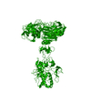 8rk4C 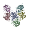 8rk5C  8rk6C  8rk7C 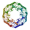 8rk9C 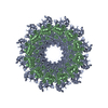 8rkaC 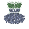 8rkbC 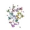 8rkcC  8rknC  8rkoC 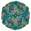 8rkxC 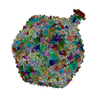 8rqeC M: map data used to model this data C: citing same article ( |
|---|---|
| Similar structure data |


-Links
 PDBj
PDBj- Assembly
| Deposited unit | 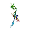
|
|---|---|
| 1 | 
|
-Components
| #1: Protein | Mass: 26983.729 Da / Num. of mol.: 1 / Source method: isolated from a natural source / Source: (natural) Pseudomonas phage JBD30 (virus) / References: UniProt: L7P801 |
|---|---|
| Has protein modification | N |
-Experimental details
-Experiment
| Experiment | Method: ELECTRON MICROSCOPY |
|---|---|
| EM experiment | Aggregation state: PARTICLE / 3D reconstruction method: single particle reconstruction |
- Sample preparation
| Component | Name: Pseudomonas phage JBD30 / Type: VIRUS Details: Phage JBD30 was propagated in P. aeruginosa strain BAA-28 and purified using CsCl gradient. Entity ID: all / Source: NATURAL | ||||||||||||||||||||
|---|---|---|---|---|---|---|---|---|---|---|---|---|---|---|---|---|---|---|---|---|---|
| Molecular weight | Value: 0.162 MDa / Experimental value: NO | ||||||||||||||||||||
| Source (natural) | Organism: Pseudomonas phage JBD30 (virus) | ||||||||||||||||||||
| Details of virus | Empty: NO / Enveloped: NO / Isolate: STRAIN / Type: VIRION | ||||||||||||||||||||
| Natural host | Organism: Pseudomonas aeruginosa / Strain: BAA-28 | ||||||||||||||||||||
| Virus shell | Name: JBD30 capsid / Diameter: 640 nm / Triangulation number (T number): 7 | ||||||||||||||||||||
| Buffer solution | pH: 8 / Details: 10 mM NaCl, 10 mM MgSO4, 50 mM Tris-HCl | ||||||||||||||||||||
| Buffer component |
| ||||||||||||||||||||
| Specimen | Embedding applied: NO / Shadowing applied: NO / Staining applied: NO / Vitrification applied: YES / Details: phage titer 10^11 PFU/ml | ||||||||||||||||||||
| Specimen support | Details: Gatan Solaris II / Grid material: COPPER / Grid mesh size: 300 divisions/in. / Grid type: Quantifoil R2/1 | ||||||||||||||||||||
| Vitrification | Instrument: FEI VITROBOT MARK IV / Cryogen name: ETHANE / Humidity: 100 % / Chamber temperature: 277.15 K Details: blotting force 0, blotting time 2s, waiting time 15s |
- Electron microscopy imaging
| Experimental equipment |  Model: Titan Krios / Image courtesy: FEI Company |
|---|---|
| Microscopy | Model: FEI TITAN KRIOS |
| Electron gun | Electron source:  FIELD EMISSION GUN / Accelerating voltage: 300 kV / Illumination mode: FLOOD BEAM FIELD EMISSION GUN / Accelerating voltage: 300 kV / Illumination mode: FLOOD BEAM |
| Electron lens | Mode: BRIGHT FIELD / Nominal magnification: 105000 X / Nominal defocus max: 1600 nm / Nominal defocus min: 600 nm / Cs: 2.7 mm / C2 aperture diameter: 50 µm / Alignment procedure: COMA FREE |
| Specimen holder | Cryogen: NITROGEN / Specimen holder model: FEI TITAN KRIOS AUTOGRID HOLDER |
| Image recording | Average exposure time: 2 sec. / Electron dose: 34 e/Å2 / Film or detector model: GATAN K3 (6k x 4k) / Num. of grids imaged: 1 / Num. of real images: 12356 |
| EM imaging optics | Energyfilter name: GIF Bioquantum / Energyfilter slit width: 10 eV |
| Image scans | Width: 5760 / Height: 4092 |
- Processing
| EM software |
| ||||||||||||||||||||||||||||||||||||||||||||||||||
|---|---|---|---|---|---|---|---|---|---|---|---|---|---|---|---|---|---|---|---|---|---|---|---|---|---|---|---|---|---|---|---|---|---|---|---|---|---|---|---|---|---|---|---|---|---|---|---|---|---|---|---|
| CTF correction | Type: PHASE FLIPPING ONLY | ||||||||||||||||||||||||||||||||||||||||||||||||||
| Particle selection | Num. of particles selected: 1780 | ||||||||||||||||||||||||||||||||||||||||||||||||||
| Symmetry | Point symmetry: C6 (6 fold cyclic) | ||||||||||||||||||||||||||||||||||||||||||||||||||
| 3D reconstruction | Resolution: 3.88 Å / Resolution method: FSC 0.143 CUT-OFF / Num. of particles: 1759 / Algorithm: FOURIER SPACE / Num. of class averages: 1 / Symmetry type: POINT | ||||||||||||||||||||||||||||||||||||||||||||||||||
| Atomic model building | Protocol: OTHER / Space: REAL | ||||||||||||||||||||||||||||||||||||||||||||||||||
| Refine LS restraints |
|
