 Movie
Movie Controller


+ Open data

- Basic information
| Entry | Database: PDB / ID: 8rk0 | |||||||||
|---|---|---|---|---|---|---|---|---|---|---|
| Title | HCV E1/E2 homodimer complex, ectodomain | |||||||||
 Components Components |
| |||||||||
 Keywords Keywords | VIRAL PROTEIN / Hepatitis C virus S52 E1 E2 Homodimer Structure | |||||||||
| Function / homology |  Function and homology information Function and homology informationhost cell mitochondrial membrane / host cell lipid droplet / symbiont-mediated transformation of host cell / symbiont-mediated suppression of host TRAF-mediated signal transduction / symbiont-mediated perturbation of host cell cycle G1/S transition checkpoint / symbiont-mediated suppression of host JAK-STAT cascade via inhibition of STAT1 activity / symbiont-mediated suppression of host cytoplasmic pattern recognition receptor signaling pathway via inhibition of MAVS activity / ribonucleoside triphosphate phosphatase activity / viral nucleocapsid / channel activity ...host cell mitochondrial membrane / host cell lipid droplet / symbiont-mediated transformation of host cell / symbiont-mediated suppression of host TRAF-mediated signal transduction / symbiont-mediated perturbation of host cell cycle G1/S transition checkpoint / symbiont-mediated suppression of host JAK-STAT cascade via inhibition of STAT1 activity / symbiont-mediated suppression of host cytoplasmic pattern recognition receptor signaling pathway via inhibition of MAVS activity / ribonucleoside triphosphate phosphatase activity / viral nucleocapsid / channel activity / monoatomic ion transmembrane transport / clathrin-dependent endocytosis of virus by host cell / RNA helicase activity / symbiont-mediated suppression of host innate immune response / host cell perinuclear region of cytoplasm / host cell endoplasmic reticulum membrane / symbiont-mediated suppression of host type I interferon-mediated signaling pathway / ribonucleoprotein complex / serine-type endopeptidase activity / symbiont-mediated activation of host autophagy / cysteine-type endopeptidase activity / viral RNA genome replication / RNA-directed RNA polymerase activity / fusion of virus membrane with host endosome membrane / viral envelope / virion attachment to host cell / host cell plasma membrane / host cell nucleus / virion membrane / structural molecule activity / proteolysis / RNA binding / zinc ion binding / ATP binding Similarity search - Function | |||||||||
| Biological species |  Hepacivirus hominis Hepacivirus hominis | |||||||||
| Method | ELECTRON MICROSCOPY / single particle reconstruction / cryo EM / Resolution: 3.38 Å | |||||||||
 Authors Authors | Augestad, E.H. / Olesen, C.H. / Groenberg, C. / Soerensen, A. / Velazquez-Moctezuma, R. / Fanalista, M. / Bukh, J. / Wang, K. / Gourdon, P. / Prentoe, J. | |||||||||
| Funding support |  Denmark, 2items Denmark, 2items
| |||||||||
 Citation Citation | Journal: Nature / Year: 2024 Title: The hepatitis C virus envelope protein complex is a dimer of heterodimers. Authors: Elias Honerød Augestad / Christina Holmboe Olesen / Christina Grønberg / Andreas Soerensen / Rodrigo Velázquez-Moctezuma / Margherita Fanalista / Jens Bukh / Kaituo Wang / Pontus Gourdon ...Authors: Elias Honerød Augestad / Christina Holmboe Olesen / Christina Grønberg / Andreas Soerensen / Rodrigo Velázquez-Moctezuma / Margherita Fanalista / Jens Bukh / Kaituo Wang / Pontus Gourdon / Jannick Prentoe /   Abstract: Fifty-eight million individuals worldwide are affected by chronic hepatitis C virus (HCV) infection, a primary driver of liver cancer for which no vaccine is available. The HCV envelope proteins E1 ...Fifty-eight million individuals worldwide are affected by chronic hepatitis C virus (HCV) infection, a primary driver of liver cancer for which no vaccine is available. The HCV envelope proteins E1 and E2 form a heterodimer (E1/E2), which is the target for neutralizing antibodies. However, the higher-order organization of these E1/E2 heterodimers, as well as that of any Hepacivirus envelope protein complex, remains unknown. Here we determined the cryo-electron microscopy structure of two E1/E2 heterodimers in a homodimeric arrangement. We reveal how the homodimer is established at the molecular level and provide insights into neutralizing antibody evasion and membrane fusion by HCV, as orchestrated by E2 motifs such as hypervariable region 1 and antigenic site 412, as well as the organization of the transmembrane helices, including two internal to E1. This study addresses long-standing questions on the higher-order oligomeric arrangement of Hepacivirus envelope proteins and provides a critical framework in the design of novel HCV vaccine antigens. | |||||||||
| History |
|
- Structure visualization
| Structure viewer | Molecule: MolmilJmol/JSmol |
|---|
- Downloads & links
-Download
| PDBx/mmCIF format | 8rk0.cif.gz | 185.4 KB | Display | PDBx/mmCIF format |
|---|---|---|---|---|
| PDB format | pdb8rk0.ent.gz | 148.2 KB | Display | PDB format |
| PDBx/mmJSON format | 8rk0.json.gz | Tree view | PDBx/mmJSON format | |
| Others |  Other downloads Other downloads |
-Validation report
| Arichive directory | https://data.pdbj.org/pub/pdb/validation_reports/rk/8rk0ftp://data.pdbj.org/pub/pdb/validation_reports/rk/8rk0 | HTTPS FTP |
|---|
-Related structure data
| Related structure data |  19254MC 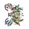 8rjjC C: citing same article ( M: map data used to model this data |
|---|---|
| Similar structure data |
-Links
 PDBj
PDBj
- Assembly
| Deposited unit | 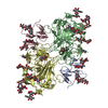
|
|---|---|
| 1 |
|
-Components
| #1: Protein | Mass: 13212.862 Da / Num. of mol.: 2 Source method: isolated from a genetically manipulated source Source: (gene. exp.) Hepacivirus hominis / Strain: S52 / Gene: E1 / Cell line (production host): HEK293 / Production host:  Homo sapiens (human) / References: UniProt: Q81424 Homo sapiens (human) / References: UniProt: Q81424#2: Protein | Mass: 36466.168 Da / Num. of mol.: 2 Source method: isolated from a genetically manipulated source Source: (gene. exp.) Hepacivirus hominis / Strain: S52 / Cell line (production host): HEK293 / Production host: Homo sapiens (human) / References: UniProt: A9YFN8#3: Polysaccharide | 2-acetamido-2-deoxy-beta-D-glucopyranose-(1-4)-2-acetamido-2-deoxy-beta-D-glucopyranose Source method: isolated from a genetically manipulated source #4: Polysaccharide | beta-D-mannopyranose-(1-4)-2-acetamido-2-deoxy-beta-D-glucopyranose-(1-4)-2-acetamido-2-deoxy-beta- ...beta-D-mannopyranose-(1-4)-2-acetamido-2-deoxy-beta-D-glucopyranose-(1-4)-2-acetamido-2-deoxy-beta-D-glucopyranose Source method: isolated from a genetically manipulated source #5: Sugar |   Type: D-saccharide, beta linking / Mass: 221.208 Da / Num. of mol.: 2 / Source method: obtained synthetically / Formula: C8H15NO6 / Feature type: SUBJECT OF INVESTIGATION Type: D-saccharide, beta linking / Mass: 221.208 Da / Num. of mol.: 2 / Source method: obtained synthetically / Formula: C8H15NO6 / Feature type: SUBJECT OF INVESTIGATIONHas ligand of interest | Y | Has protein modification | Y | |
|---|
-Experimental details
-Experiment
| Experiment | Method: ELECTRON MICROSCOPY |
|---|---|
| EM experiment | Aggregation state: PARTICLE / 3D reconstruction method: single particle reconstruction |
- Sample preparation
| Component | Name: HCV S52 E1E2 / Type: COMPLEX / Entity ID: #1-#2 / Source: RECOMBINANT |
|---|---|
| Molecular weight | Value: 0.2 MDa / Experimental value: NO |
| Source (natural) | Organism: Hepacivirus hominis |
| Source (recombinant) | Organism: Homo sapiens (human) / Cell: HEK293 |
| Buffer solution | pH: 7.5 |
| Specimen | Embedding applied: NO / Shadowing applied: NO / Staining applied: NO / Vitrification applied: YES |
| Vitrification | Instrument: FEI VITROBOT MARK IV / Cryogen name: ETHANE / Humidity: 100 % / Chamber temperature: 277 K |
- Electron microscopy imaging
| Experimental equipment |  Model: Titan Krios / Image courtesy: FEI Company |
|---|---|
| Microscopy | Model: FEI TITAN KRIOS |
| Electron gun | Electron source:  FIELD EMISSION GUN / Accelerating voltage: 300 kV / Illumination mode: OTHER FIELD EMISSION GUN / Accelerating voltage: 300 kV / Illumination mode: OTHER |
| Electron lens | Mode: BRIGHT FIELD / Nominal defocus max: 2500 nm / Nominal defocus min: 1000 nm / Cs: 2.7 mm / C2 aperture diameter: 100 µm / Alignment procedure: BASIC |
| Specimen holder | Cryogen: NITROGEN / Specimen holder model: FEI TITAN KRIOS AUTOGRID HOLDER |
| Image recording | Electron dose: 50 e/Å2 / Film or detector model: GATAN K3 (6k x 4k) / Num. of grids imaged: 1 / Num. of real images: 13555 |
| Image scans | Width: 4092 / Height: 5760 |
- Processing
| EM software |
| ||||||||||||||||||||||||||||||||
|---|---|---|---|---|---|---|---|---|---|---|---|---|---|---|---|---|---|---|---|---|---|---|---|---|---|---|---|---|---|---|---|---|---|
| CTF correction | Type: PHASE FLIPPING AND AMPLITUDE CORRECTION | ||||||||||||||||||||||||||||||||
| 3D reconstruction | Resolution: 3.38 Å / Resolution method: FSC 0.143 CUT-OFF / Num. of particles: 105033 / Symmetry type: POINT | ||||||||||||||||||||||||||||||||
| Atomic model building | B value: 100 / Protocol: FLEXIBLE FIT / Space: REAL | ||||||||||||||||||||||||||||||||
| Atomic model building | Source name: AlphaFold / Type: in silico model | ||||||||||||||||||||||||||||||||
| Refine LS restraints |
|
