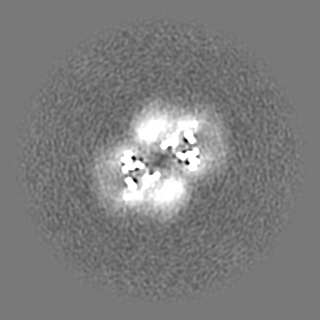
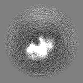
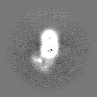
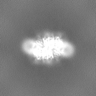
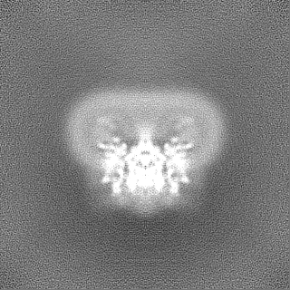
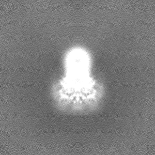
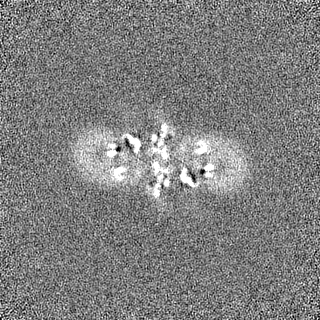
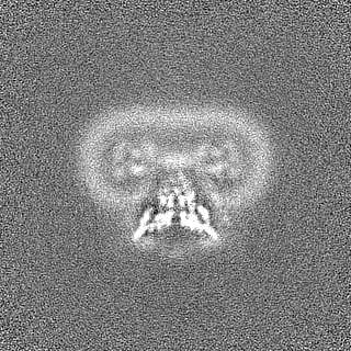
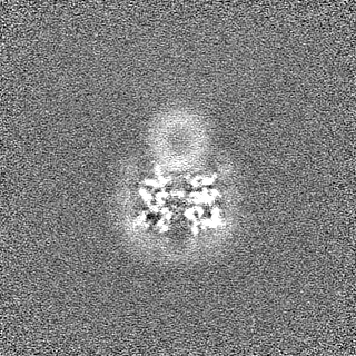
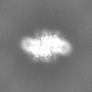
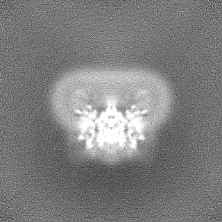
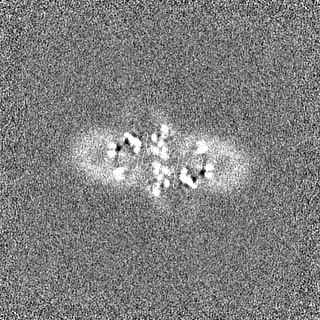
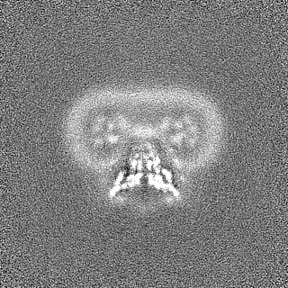
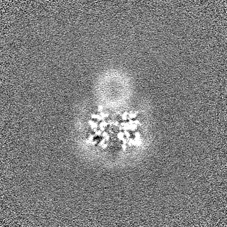
Journal: Nature / Year: 2024 Title: The hepatitis C virus envelope protein complex is a dimer of heterodimers. Authors: Elias Honerød Augestad / Christina Holmboe Olesen / Christina Grønberg / Andreas Soerensen / Rodrigo Velázquez-Moctezuma / Margherita Fanalista / Jens Bukh / Kaituo Wang / Pontus Gourdon ...Authors: Elias Honerød Augestad / Christina Holmboe Olesen / Christina Grønberg / Andreas Soerensen / Rodrigo Velázquez-Moctezuma / Margherita Fanalista / Jens Bukh / Kaituo Wang / Pontus Gourdon / Jannick Prentoe / Abstract: Fifty-eight million individuals worldwide are affected by chronic hepatitis C virus (HCV) infection, a primary driver of liver cancer for which no vaccine is available. The HCV envelope proteins E1 ...Fifty-eight million individuals worldwide are affected by chronic hepatitis C virus (HCV) infection, a primary driver of liver cancer for which no vaccine is available. The HCV envelope proteins E1 and E2 form a heterodimer (E1/E2), which is the target for neutralizing antibodies. However, the higher-order organization of these E1/E2 heterodimers, as well as that of any Hepacivirus envelope protein complex, remains unknown. Here we determined the cryo-electron microscopy structure of two E1/E2 heterodimers in a homodimeric arrangement. We reveal how the homodimer is established at the molecular level and provide insights into neutralizing antibody evasion and membrane fusion by HCV, as orchestrated by E2 motifs such as hypervariable region 1 and antigenic site 412, as well as the organization of the transmembrane helices, including two internal to E1. This study addresses long-standing questions on the higher-order oligomeric arrangement of Hepacivirus envelope proteins and provides a critical framework in the design of novel HCV vaccine antigens.
In the structure databanks used in Yorodumi, some data are registered as the other names, "COVID-19 virus" and "2019-nCoV". Here are the details of the virus and the list of structure data.
Jan 31, 2019. EMDB accession codes are about to change! (news from PDBe EMDB page)
EMDB accession codes are about to change! (news from PDBe EMDB page)
The allocation of 4 digits for EMDB accession codes will soon come to an end. Whilst these codes will remain in use, new EMDB accession codes will include an additional digit and will expand incrementally as the available range of codes is exhausted. The current 4-digit format prefixed with “EMD-” (i.e. EMD-XXXX) will advance to a 5-digit format (i.e. EMD-XXXXX), and so on. It is currently estimated that the 4-digit codes will be depleted around Spring 2019, at which point the 5-digit format will come into force.
The EM Navigator/Yorodumi systems omit the EMD- prefix.
Related info.:Q: What is EMD? / ID/Accession-code notation in Yorodumi/EM Navigator
Yorodumi is a browser for structure data from EMDB, PDB, SASBDB, etc.
This page is also the successor to EM Navigator detail page, and also detail information page/front-end page for Omokage search.
The word "yorodu" (or yorozu) is an old Japanese word meaning "ten thousand". "mi" (miru) is to see.
Related info.:EMDB / PDB / SASBDB / Comparison of 3 databanks / Yorodumi Search / Aug 31, 2016. New EM Navigator & Yorodumi / Yorodumi Papers / Jmol/JSmol / Function and homology information / Changes in new EM Navigator and Yorodumi
 Movie
Movie Controller


 Open data
 Basic information
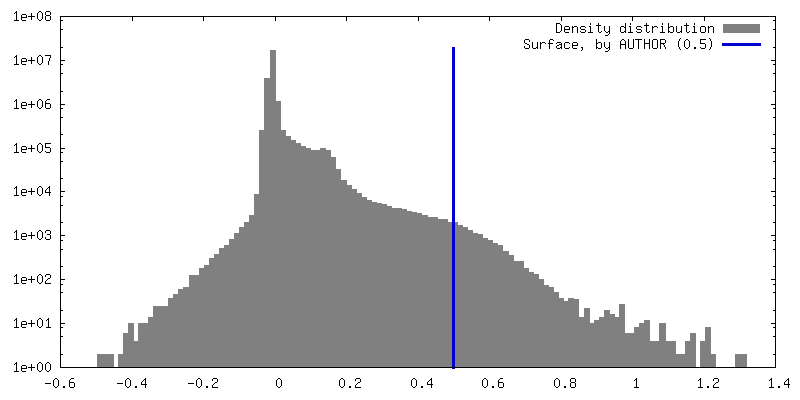
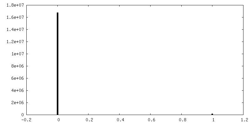
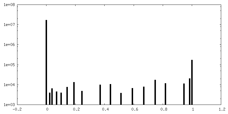
 Map data
 Sample
 Keywords
 Function and homology information
 Hepacivirus hominis
 Authors
 Denmark, 2 items
  Citation

 Structure visualization
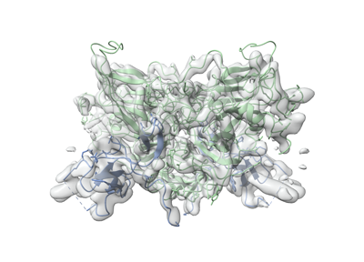
 Downloads & links
 emd_19254.png
 http://ftp.pdbj.org/pub/emdb/structures/EMD-19254

 Z (Sec.)
 Y (Row.)
 X (Col.)


 Sample components
 Homo sapiens (human)
 Processing
 Electron microscopy
 FIELD EMISSION GUN
