 Movie
Movie Controller

[English] 日本語
 Yorodumi
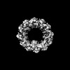
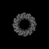
- PDB-8rgx: Cryo-EM structure of the Bacterial Proteasome Activator Bpa of My... -
+ Open data

- Basic information
| Entry | Database: PDB / ID: 8rgx | ||||||
|---|---|---|---|---|---|---|---|
| Title | Cryo-EM structure of the Bacterial Proteasome Activator Bpa of Mycobacterium tuberculosis | ||||||
 Components Components | Bacterial proteasome activator | ||||||
 Keywords Keywords | CHAPERONE / protein degradation / quality control / proteasomal activator / mycobacteria | ||||||
| Function / homology | Bacterial proteasome activator / Bacterial proteasome activator / proteasome accessory complex / positive regulation of proteasomal protein catabolic process / proteasome binding / peptidoglycan-based cell wall / protein homooligomerization / plasma membrane / Bacterial proteasome activator Function and homology information Function and homology information | ||||||
| Biological species |  Mycobacterium tuberculosis H37Rv (bacteria) Mycobacterium tuberculosis H37Rv (bacteria) | ||||||
| Method | ELECTRON MICROSCOPY / single particle reconstruction / cryo EM / Resolution: 3.2 Å | ||||||
 Authors Authors | Zdanowicz, R. / von Rosen, T. / Afanasyev, P. / Boehringer, D. / Glockshuber, R. / Weber-Ban, E. | ||||||
| Funding support |  Switzerland, 1items Switzerland, 1items
| ||||||
 Citation Citation | Journal: Nat Commun / Year: 2025 Title: Substrates bind to residues lining the ring of asymmetrically engaged bacterial proteasome activator Bpa. Authors: Tatjana von Rosen / Rafal Zdanowicz / Yasser El Hadeg / Pavel Afanasyev / Daniel Boehringer / Alexander Leitner / Rudi Glockshuber / Eilika Weber-Ban /  Abstract: Mycobacteria harbor a proteasome that was acquired by Actinobacteria through horizontal gene transfer and that supports the persistence of the human pathogen Mycobacterium tuberculosis within host ...Mycobacteria harbor a proteasome that was acquired by Actinobacteria through horizontal gene transfer and that supports the persistence of the human pathogen Mycobacterium tuberculosis within host macrophages. The core particle of the proteasome (20S CP) associates with ring-shaped activator complexes to degrade protein substrates. One of these is the bacterial proteasome activator Bpa that stimulates the ATP-independent proteasomal degradation of the heat shock repressor HspR. In this study, we determine the cryogenic electron microscopy 3D reconstruction of the complex between Bpa and its natural substrate HspR at 4.1 Å global resolution. The resulting maps allow us to identify regions of Bpa that interact with HspR. Using structure-guided site-directed mutagenesis and in vitro biochemical assays, we confirm the importance of the identified residues for Bpa-mediated substrate recruitment and subsequent proteasomal degradation. Additionally, we show that the dodecameric Bpa ring associates asymmetrically with the heptameric α-rings of the 20S CP, adopting a conformation resembling a hinged lid, while still engaging all seven docking sites on the proteasome. | ||||||
| History |
|
- Structure visualization
| Structure viewer | Molecule: MolmilJmol/JSmol |
|---|
- Downloads & links
-Download
| PDBx/mmCIF format | 8rgx.cif.gz | 228.2 KB | Display | PDBx/mmCIF format |
|---|---|---|---|---|
| PDB format | pdb8rgx.ent.gz | 179.8 KB | Display | PDB format |
| PDBx/mmJSON format | 8rgx.json.gz | Tree view | PDBx/mmJSON format | |
| Others |  Other downloads Other downloads |
-Validation report
| Arichive directory | https://data.pdbj.org/pub/pdb/validation_reports/rg/8rgxftp://data.pdbj.org/pub/pdb/validation_reports/rg/8rgx | HTTPS FTP |
|---|
-Related structure data
| Related structure data | 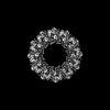 19162MC M: map data used to model this data C: citing same article ( |
|---|---|
| Similar structure data |
-Links
 PDBj
PDBj
- Assembly
| Deposited unit | 
|
|---|---|
| 1 |
|
-Components
| #1: Protein | Mass: 19923.311 Da / Num. of mol.: 12 Source method: isolated from a genetically manipulated source Source: (gene. exp.) Mycobacterium tuberculosis H37Rv (bacteria)Gene: bpa / Production host: Has protein modification | N | |
|---|
-Experimental details
-Experiment
| Experiment | Method: ELECTRON MICROSCOPY |
|---|---|
| EM experiment | Aggregation state: PARTICLE / 3D reconstruction method: single particle reconstruction |
- Sample preparation
| Component | Name: Bacterial Proteasome Activator Bpa of Mycobacterium tuberculosis Type: COMPLEX / Entity ID: all / Source: RECOMBINANT |
|---|---|
| Molecular weight | Experimental value: NO |
| Source (natural) | Organism: Mycobacterium tuberculosis H37Rv (bacteria) |
| Source (recombinant) | Organism: |
| Buffer solution | pH: 7.5 |
| Specimen | Embedding applied: NO / Shadowing applied: NO / Staining applied: NO / Vitrification applied: YES |
| Specimen support | Grid material: COPPER / Grid type: Quantifoil R2/2 |
| Vitrification | Instrument: FEI VITROBOT MARK IV / Cryogen name: ETHANE-PROPANE |
- Electron microscopy imaging
| Experimental equipment |  Model: Titan Krios / Image courtesy: FEI Company |
|---|---|
| Microscopy | Model: FEI TITAN KRIOS |
| Electron gun | Electron source:  FIELD EMISSION GUN / Accelerating voltage: 300 kV / Illumination mode: FLOOD BEAM FIELD EMISSION GUN / Accelerating voltage: 300 kV / Illumination mode: FLOOD BEAM |
| Electron lens | Mode: BRIGHT FIELD / Nominal defocus max: 2600 nm / Nominal defocus min: 1200 nm / Cs: 2.7 mm |
| Image recording | Electron dose: 80 e/Å2 / Film or detector model: GATAN K3 (6k x 4k) |
- Processing
| EM software |
| |||||||||
|---|---|---|---|---|---|---|---|---|---|---|
| CTF correction | Type: PHASE FLIPPING AND AMPLITUDE CORRECTION | |||||||||
| Symmetry | Point symmetry: C12 (12 fold cyclic) | |||||||||
| 3D reconstruction | Resolution: 3.2 Å / Resolution method: FSC 0.143 CUT-OFF / Num. of particles: 229813 / Symmetry type: POINT | |||||||||
| Atomic model building | PDB-ID: 5LFJ Accession code: 5LFJ / Details: Bacterial proteasome activator Bpa / Source name: PDB / Type: experimental model |

