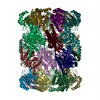
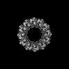
Journal: Nat Commun / Year: 2025 Title: Substrates bind to residues lining the ring of asymmetrically engaged bacterial proteasome activator Bpa. Authors: Tatjana von Rosen / Rafal Zdanowicz / Yasser El Hadeg / Pavel Afanasyev / Daniel Boehringer / Alexander Leitner / Rudi Glockshuber / Eilika Weber-Ban / Abstract: Mycobacteria harbor a proteasome that was acquired by Actinobacteria through horizontal gene transfer and that supports the persistence of the human pathogen Mycobacterium tuberculosis within host ...Mycobacteria harbor a proteasome that was acquired by Actinobacteria through horizontal gene transfer and that supports the persistence of the human pathogen Mycobacterium tuberculosis within host macrophages. The core particle of the proteasome (20S CP) associates with ring-shaped activator complexes to degrade protein substrates. One of these is the bacterial proteasome activator Bpa that stimulates the ATP-independent proteasomal degradation of the heat shock repressor HspR. In this study, we determine the cryogenic electron microscopy 3D reconstruction of the complex between Bpa and its natural substrate HspR at 4.1 Å global resolution. The resulting maps allow us to identify regions of Bpa that interact with HspR. Using structure-guided site-directed mutagenesis and in vitro biochemical assays, we confirm the importance of the identified residues for Bpa-mediated substrate recruitment and subsequent proteasomal degradation. Additionally, we show that the dodecameric Bpa ring associates asymmetrically with the heptameric α-rings of the 20S CP, adopting a conformation resembling a hinged lid, while still engaging all seven docking sites on the proteasome.
Entire : Complex of the 20S proteasome with bacterial proteasome activator...
Entire
Name: Complex of the 20S proteasome with bacterial proteasome activator Bpa from M. tuberculosis
Components
Complex: Complex of the 20S proteasome with bacterial proteasome activator Bpa from M. tuberculosis
Protein or peptide: Proteasome subunit alpha
Protein or peptide: Proteasome subunit beta
Protein or peptide: Bacterial proteasome activator
-
Supramolecule #1: Complex of the 20S proteasome with bacterial proteasome activator...
Supramolecule
Name: Complex of the 20S proteasome with bacterial proteasome activator Bpa from M. tuberculosis type: complex / ID: 1 / Parent: 0 / Macromolecule list: all
In the structure databanks used in Yorodumi, some data are registered as the other names, "COVID-19 virus" and "2019-nCoV". Here are the details of the virus and the list of structure data.
Jan 31, 2019. EMDB accession codes are about to change! (news from PDBe EMDB page)
EMDB accession codes are about to change! (news from PDBe EMDB page)
The allocation of 4 digits for EMDB accession codes will soon come to an end. Whilst these codes will remain in use, new EMDB accession codes will include an additional digit and will expand incrementally as the available range of codes is exhausted. The current 4-digit format prefixed with “EMD-” (i.e. EMD-XXXX) will advance to a 5-digit format (i.e. EMD-XXXXX), and so on. It is currently estimated that the 4-digit codes will be depleted around Spring 2019, at which point the 5-digit format will come into force.
The EM Navigator/Yorodumi systems omit the EMD- prefix.
Related info.:Q: What is EMD? / ID/Accession-code notation in Yorodumi/EM Navigator
Yorodumi is a browser for structure data from EMDB, PDB, SASBDB, etc.
This page is also the successor to EM Navigator detail page, and also detail information page/front-end page for Omokage search.
The word "yorodu" (or yorozu) is an old Japanese word meaning "ten thousand". "mi" (miru) is to see.
Related info.:EMDB / PDB / SASBDB / Comparison of 3 databanks / Yorodumi Search / Aug 31, 2016. New EM Navigator & Yorodumi / Yorodumi Papers / Jmol/JSmol / Function and homology information / Changes in new EM Navigator and Yorodumi
 Movie
Movie Controller

 Yorodumi
 Open data
 Basic information
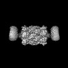
 Map data
 Sample
 Keywords
 Function and homology information
 Mycobacterium tuberculosis H37Rv (bacteria)
 Authors
 Switzerland, 1 items
  Citation
 Structure visualization
 Downloads & links
 emd_19150.png
 http://ftp.pdbj.org/pub/emdb/structures/EMD-19150
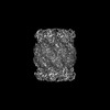


 Z (Sec.)
 Y (Row.)
 X (Col.)


 Sample components
 Processing
 Electron microscopy
 FIELD EMISSION GUN