 Movie
Movie Controller

[English] 日本語
 Yorodumi
- PDB-8rg1: Soluble glucose dehydrogenase from acinetobacter calcoaceticus - ... -
+ Open data

- Basic information
| Entry | Database: PDB / ID: 8rg1 | ||||||
|---|---|---|---|---|---|---|---|
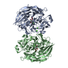
| Title | Soluble glucose dehydrogenase from acinetobacter calcoaceticus - wild type pH8 | ||||||
 Components Components | Quinoprotein glucose dehydrogenase B | ||||||
 Keywords Keywords | OXIDOREDUCTASE / Wild type / PQQ | ||||||
| Function / homology |  Function and homology information Function and homology informationglucose 1-dehydrogenase (PQQ, quinone) / quinoprotein glucose dehydrogenase activity / metal ion binding Similarity search - Function | ||||||
| Biological species |  Acinetobacter calcoaceticus (bacteria) Acinetobacter calcoaceticus (bacteria) | ||||||
| Method |  X-RAY DIFFRACTION / SYNCHROTRON / MOLECULAR REPLACEMENT / Resolution: 1.19 Å X-RAY DIFFRACTION / SYNCHROTRON / MOLECULAR REPLACEMENT / Resolution: 1.19 Å | ||||||
 Authors Authors | Lublin, V. / Chavas, L. / Stines-Chaumeil, C. / Kauffmann, B. / Giraud, M.F. / Thompson, A. | ||||||
| Funding support |  France, 1items France, 1items
| ||||||
 Citation Citation | Journal: Biosci.Rep. / Year: 2024 Title: Does Acinetobacter calcoaceticus glucose dehydrogenase produce self-damaging H2O2? Authors: Lublin, V. / Kauffmann, B. / Engilberge, S. / Durola, F. / Gounel, S. / Bichon, S. / Jean, C. / Mano, N. / Giraud, M.F. / Chavas, L.M.G.H. / Thureau, A. / Thompson, A. / Stines-Chaumeil, C. #1: Journal: Acta Crystallogr., Sect. D: Biol. Crystallogr. / Year: 2012Title: Towards automated crystallographic structure refinement with phenix.refine. Authors: Afonine, P.V. | ||||||
| History |
|
- Structure visualization
| Structure viewer | Molecule: MolmilJmol/JSmol |
|---|
- Downloads & links
-Download
| PDBx/mmCIF format | 8rg1.cif.gz | 623.5 KB | Display | PDBx/mmCIF format |
|---|---|---|---|---|
| PDB format | pdb8rg1.ent.gz | Display | PDB format | |
| PDBx/mmJSON format | 8rg1.json.gz | Tree view | PDBx/mmJSON format | |
| Others |  Other downloads Other downloads |
-Validation report
| Summary document | 8rg1_validation.pdf.gz | 1.2 MB | Display | wwPDB validaton report |
|---|---|---|---|---|
| Full document | 8rg1_full_validation.pdf.gz | 1.2 MB | Display | |
| Data in XML | 8rg1_validation.xml.gz | 47 KB | Display | |
| Data in CIF | 8rg1_validation.cif.gz | 74.8 KB | Display | |
| Arichive directory | https://data.pdbj.org/pub/pdb/validation_reports/rg/8rg1ftp://data.pdbj.org/pub/pdb/validation_reports/rg/8rg1 | HTTPS FTP |
-Related structure data

-Links
 PDBj
PDBj


- Assembly
| Deposited unit | 
| ||||||||||||
|---|---|---|---|---|---|---|---|---|---|---|---|---|---|
| 1 |
| ||||||||||||
| Unit cell |
|
-Components
| #1: Protein | Mass: 50293.207 Da / Num. of mol.: 2 Source method: isolated from a genetically manipulated source Source: (gene. exp.) Acinetobacter calcoaceticus (bacteria) / Gene: gdhB / Production host: #2: Chemical | Mass: 364.221 Da / Num. of mol.: 2 / Source method: obtained synthetically / Formula: C14H8N2O10 / Feature type: SUBJECT OF INVESTIGATION #3: Chemical | ChemComp-CA /   Mass: 40.078 Da / Num. of mol.: 6 / Source method: obtained synthetically / Formula: Ca Mass: 40.078 Da / Num. of mol.: 6 / Source method: obtained synthetically / Formula: Ca#4: Chemical |   Mass: 6.941 Da / Num. of mol.: 2 / Source method: obtained synthetically / Formula: Li Mass: 6.941 Da / Num. of mol.: 2 / Source method: obtained synthetically / Formula: Li#5: Water | ChemComp-HOH / |  Mass: 18.015 Da / Num. of mol.: 1383 / Source method: isolated from a natural source / Formula: H2O Mass: 18.015 Da / Num. of mol.: 1383 / Source method: isolated from a natural source / Formula: H2OHas ligand of interest | Y | Has protein modification | Y | |
|---|
-Experimental details
-Experiment
| Experiment | Method: X-RAY DIFFRACTION / Number of used crystals: 1 |
|---|
- Sample preparation
| Crystal | Density Matthews: 2.27 Å3/Da / Density % sol: 45.84 % |
|---|---|
| Crystal grow | Temperature: 293 K / Method: vapor diffusion, sitting drop / pH: 8 / Details: PEG 6000, 100 mM TRIS pH8, 2 mM ZnCl2 |
-Data collection
| Diffraction | Mean temperature: 100 K / Serial crystal experiment: N |
|---|---|
| Diffraction source | Source: SYNCHROTRON / Site: ESRF / Beamline: BM07 / Wavelength: 0.98 Å |
| Detector | Type: DECTRIS PILATUS 6M / Detector: PIXEL / Date: Feb 6, 2023 |
| Radiation | Protocol: SINGLE WAVELENGTH / Monochromatic (M) / Laue (L): M / Scattering type: x-ray |
| Radiation wavelength | Wavelength: 0.98 Å / Relative weight: 1 |
| Reflection | Resolution: 1.18→50 Å / Num. obs: 199471 / % possible obs: 98.7 % / Redundancy: 6.7 % / Biso Wilson estimate: 12.49 Å2 / CC1/2: 0.999 / Net I/σ(I): 16.5 |
| Reflection shell | Resolution: 1.19→1.233 Å / Redundancy: 2 % / Rmerge(I) obs: 0.42 / Num. unique obs: 1815 / CC1/2: 0.648 / CC star: 0.887 / Rpim(I) all: 0.42 / Rrim(I) all: 0.59 / % possible all: 6.35 |
- Processing
| Software |
| |||||||||||||||||||||||||||||||||||||||||||||||||||||||||||||||||||||||||||||||||||||||||||||||||||||||||
|---|---|---|---|---|---|---|---|---|---|---|---|---|---|---|---|---|---|---|---|---|---|---|---|---|---|---|---|---|---|---|---|---|---|---|---|---|---|---|---|---|---|---|---|---|---|---|---|---|---|---|---|---|---|---|---|---|---|---|---|---|---|---|---|---|---|---|---|---|---|---|---|---|---|---|---|---|---|---|---|---|---|---|---|---|---|---|---|---|---|---|---|---|---|---|---|---|---|---|---|---|---|---|---|---|---|---|
| Refinement | Method to determine structure: MOLECULAR REPLACEMENT / Resolution: 1.19→42.56 Å / SU ML: 0.109 / Cross valid method: FREE R-VALUE / σ(F): 1.34 / Phase error: 22.971 Stereochemistry target values: GeoStd + Monomer Library + CDL v1.2
| |||||||||||||||||||||||||||||||||||||||||||||||||||||||||||||||||||||||||||||||||||||||||||||||||||||||||
| Solvent computation | Shrinkage radii: 0.9 Å / VDW probe radii: 1.11 Å / Solvent model: FLAT BULK SOLVENT MODEL | |||||||||||||||||||||||||||||||||||||||||||||||||||||||||||||||||||||||||||||||||||||||||||||||||||||||||
| Displacement parameters | Biso mean: 19.64 Å2 | |||||||||||||||||||||||||||||||||||||||||||||||||||||||||||||||||||||||||||||||||||||||||||||||||||||||||
| Refinement step | Cycle: LAST / Resolution: 1.19→42.56 Å
| |||||||||||||||||||||||||||||||||||||||||||||||||||||||||||||||||||||||||||||||||||||||||||||||||||||||||
| Refine LS restraints |
| |||||||||||||||||||||||||||||||||||||||||||||||||||||||||||||||||||||||||||||||||||||||||||||||||||||||||
| LS refinement shell |
|
