 Movie
Movie Controller

[English] 日本語
 Yorodumi
- PDB-8rfk: Soluble glucose dehydrogenase from acinetobacter calcoaceticus - ... -
+ Open data

- Basic information
| Entry | Database: PDB / ID: 8rfk | ||||||
|---|---|---|---|---|---|---|---|
| Title | Soluble glucose dehydrogenase from acinetobacter calcoaceticus - single mutant pH8 | ||||||
 Components Components | Quinoprotein glucose dehydrogenase B | ||||||
 Keywords Keywords | OXIDOREDUCTASE / Double point mutation | ||||||
| Function / homology |  Function and homology information Function and homology informationglucose 1-dehydrogenase (PQQ, quinone) / quinoprotein glucose dehydrogenase activity / metal ion binding Similarity search - Function | ||||||
| Biological species |  Acinetobacter calcoaceticus (bacteria) Acinetobacter calcoaceticus (bacteria) | ||||||
| Method |  X-RAY DIFFRACTION / SYNCHROTRON / MOLECULAR REPLACEMENT / Resolution: 1.56 Å X-RAY DIFFRACTION / SYNCHROTRON / MOLECULAR REPLACEMENT / Resolution: 1.56 Å | ||||||
 Authors Authors | Lublin, V. / Chavas, L. / Stines-Chaumeil, C. / Kauffmann, B. / Giraud, M.F. / Thompson, A. | ||||||
| Funding support |  France, 1items France, 1items
| ||||||
 Citation Citation | Journal: Biosci.Rep. / Year: 2024 Title: Does Acinetobacter calcoaceticus glucose dehydrogenase produce self-damaging H2O2? Authors: Lublin, V. / Kauffmann, B. / Engilberge, S. / Durola, F. / Gounel, S. / Bichon, S. / Jean, C. / Mano, N. / Giraud, M.F. / Chavas, L.M.G.H. / Thureau, A. / Thompson, A. / Stines-Chaumeil, C. | ||||||
| History |
|
- Structure visualization
| Structure viewer | Molecule: MolmilJmol/JSmol |
|---|
- Downloads & links
-Download
| PDBx/mmCIF format | 8rfk.cif.gz | 458.3 KB | Display | PDBx/mmCIF format |
|---|---|---|---|---|
| PDB format | pdb8rfk.ent.gz | Display | PDB format | |
| PDBx/mmJSON format | 8rfk.json.gz | Tree view | PDBx/mmJSON format | |
| Others |  Other downloads Other downloads |
-Validation report
| Summary document | 8rfk_validation.pdf.gz | 1.2 MB | Display | wwPDB validaton report |
|---|---|---|---|---|
| Full document | 8rfk_full_validation.pdf.gz | 1.2 MB | Display | |
| Data in XML | 8rfk_validation.xml.gz | 45.8 KB | Display | |
| Data in CIF | 8rfk_validation.cif.gz | 72.5 KB | Display | |
| Arichive directory | https://data.pdbj.org/pub/pdb/validation_reports/rf/8rfkftp://data.pdbj.org/pub/pdb/validation_reports/rf/8rfk | HTTPS FTP |
-Related structure data


-Links
 PDBj
PDBj


- Assembly
| Deposited unit | 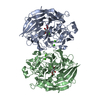
| ||||||||||||
|---|---|---|---|---|---|---|---|---|---|---|---|---|---|
| 1 |
| ||||||||||||
| Unit cell |
|
-Components
| #1: Protein | Mass: 50277.207 Da / Num. of mol.: 2 / Mutation: D143E,Y343F Source method: isolated from a genetically manipulated source Source: (gene. exp.) Acinetobacter calcoaceticus (bacteria) / Gene: gdhB / Production host: #2: Chemical | ChemComp-CA /   Mass: 40.078 Da / Num. of mol.: 6 / Source method: obtained synthetically / Formula: Ca Mass: 40.078 Da / Num. of mol.: 6 / Source method: obtained synthetically / Formula: Ca#3: Chemical | Mass: 364.221 Da / Num. of mol.: 2 / Source method: obtained synthetically / Formula: C14H8N2O10 / Feature type: SUBJECT OF INVESTIGATION #4: Water | ChemComp-HOH / |  Mass: 18.015 Da / Num. of mol.: 1300 / Source method: isolated from a natural source / Formula: H2O Mass: 18.015 Da / Num. of mol.: 1300 / Source method: isolated from a natural source / Formula: H2OHas ligand of interest | Y | Has protein modification | Y | |
|---|
-Experimental details
-Experiment
| Experiment | Method: X-RAY DIFFRACTION / Number of used crystals: 1 |
|---|
- Sample preparation
| Crystal | Density Matthews: 2.33 Å3/Da / Density % sol: 47.23 % |
|---|---|
| Crystal grow | Temperature: 300 K / Method: vapor diffusion, sitting drop / pH: 8 / Details: PEG 6000, TRIS, Lithium chloride |
-Data collection
| Diffraction | Mean temperature: 100 K / Serial crystal experiment: N |
|---|---|
| Diffraction source | Source: SYNCHROTRON / Site: SOLEIL / Beamline: PROXIMA 1 / Wavelength: 0.98 Å |
| Detector | Type: DECTRIS EIGER X 16M / Detector: PIXEL / Date: Apr 8, 2022 |
| Radiation | Protocol: SINGLE WAVELENGTH / Monochromatic (M) / Laue (L): M / Scattering type: x-ray |
| Radiation wavelength | Wavelength: 0.98 Å / Relative weight: 1 |
| Reflection | Resolution: 1.54→46.5 Å / Num. obs: 131392 / % possible obs: 97.7 % / Redundancy: 6.4 % / Biso Wilson estimate: 16.41 Å2 / CC1/2: 0.998 / Rrim(I) all: 0.132 / Net I/σ(I): 10.7 |
| Reflection shell | Resolution: 1.54→1.63 Å / Rmerge(I) obs: 1 / Mean I/σ(I) obs: 0.58 / Num. unique obs: 18670 / CC1/2: 0.36 / Rrim(I) all: 1.59 / % possible all: 86 |
- Processing
| Software |
| |||||||||||||||||||||||||||||||||||||||||||||||||||||||||||||||||||||||||||||||||||||||||||||||||||||||||
|---|---|---|---|---|---|---|---|---|---|---|---|---|---|---|---|---|---|---|---|---|---|---|---|---|---|---|---|---|---|---|---|---|---|---|---|---|---|---|---|---|---|---|---|---|---|---|---|---|---|---|---|---|---|---|---|---|---|---|---|---|---|---|---|---|---|---|---|---|---|---|---|---|---|---|---|---|---|---|---|---|---|---|---|---|---|---|---|---|---|---|---|---|---|---|---|---|---|---|---|---|---|---|---|---|---|---|
| Refinement | Method to determine structure: MOLECULAR REPLACEMENT / Resolution: 1.56→42.73 Å / SU ML: 0.1354 / Cross valid method: FREE R-VALUE / σ(F): 1.35 / Phase error: 21.8244 Stereochemistry target values: GeoStd + Monomer Library + CDL v1.2
| |||||||||||||||||||||||||||||||||||||||||||||||||||||||||||||||||||||||||||||||||||||||||||||||||||||||||
| Solvent computation | Shrinkage radii: 0.9 Å / VDW probe radii: 1.1 Å / Solvent model: FLAT BULK SOLVENT MODEL | |||||||||||||||||||||||||||||||||||||||||||||||||||||||||||||||||||||||||||||||||||||||||||||||||||||||||
| Displacement parameters | Biso mean: 21.61 Å2 | |||||||||||||||||||||||||||||||||||||||||||||||||||||||||||||||||||||||||||||||||||||||||||||||||||||||||
| Refinement step | Cycle: LAST / Resolution: 1.56→42.73 Å
| |||||||||||||||||||||||||||||||||||||||||||||||||||||||||||||||||||||||||||||||||||||||||||||||||||||||||
| Refine LS restraints |
| |||||||||||||||||||||||||||||||||||||||||||||||||||||||||||||||||||||||||||||||||||||||||||||||||||||||||
| LS refinement shell |
|
