Movie
Movie Controller


+ Open data

- Basic information
| Entry | Database: PDB / ID: 8r7n | ||||||
|---|---|---|---|---|---|---|---|
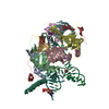
| Title | Overall structure of the U11 snRNP | ||||||
 Components Components |
| ||||||
 Keywords Keywords | SPLICING / minor spliceosome / U11 snRNP / RNA-protein complex | ||||||
| Function / homology |  Function and homology information Function and homology informationU2 snRNP binding / snRNA binding / U7 snRNA binding / histone pre-mRNA DCP binding / U7 snRNP / histone pre-mRNA 3'end processing complex / SLBP independent Processing of Histone Pre-mRNAs / SLBP Dependent Processing of Replication-Dependent Histone Pre-mRNAs / 7-methylguanosine cap hypermethylation / U12-type spliceosomal complex ...U2 snRNP binding / snRNA binding / U7 snRNA binding / histone pre-mRNA DCP binding / U7 snRNP / histone pre-mRNA 3'end processing complex / SLBP independent Processing of Histone Pre-mRNAs / SLBP Dependent Processing of Replication-Dependent Histone Pre-mRNAs / 7-methylguanosine cap hypermethylation / U12-type spliceosomal complex / protein methylation / U1 snRNP binding / pICln-Sm protein complex / methylosome / small nuclear ribonucleoprotein complex / SMN-Sm protein complex / spliceosomal tri-snRNP complex / P granule / snRNP binding / commitment complex / U2-type precatalytic spliceosome / U2-type prespliceosome assembly / U2-type catalytic step 2 spliceosome / U2-type spliceosomal complex / telomerase holoenzyme complex / telomerase RNA binding / U1 snRNP / U2 snRNP / RNA Polymerase II Transcription Termination / U4 snRNP / U2-type prespliceosome / precatalytic spliceosome / mRNA Splicing - Minor Pathway / spliceosomal complex assembly / U5 snRNP / spliceosomal snRNP assembly / U4/U6 x U5 tri-snRNP complex / intercellular bridge / catalytic step 2 spliceosome / mRNA Splicing - Major Pathway / RNA splicing / response to glucocorticoid / spliceosomal complex / mRNA splicing, via spliceosome / mRNA processing / snRNP Assembly / SARS-CoV-2 modulates host translation machinery / nuclear body / mRNA binding / apoptotic process / nucleolus / enzyme binding / RNA binding / extracellular exosome / zinc ion binding / nucleoplasm / nucleus / cytosol / cytoplasm Similarity search - Function | ||||||
| Biological species |  Homo sapiens (human) Homo sapiens (human) | ||||||
| Method | ELECTRON MICROSCOPY / single particle reconstruction / cryo EM / Resolution: 3.4 Å | ||||||
 Authors Authors | Zhao, J. / Galej, W.P. | ||||||
| Funding support | European Union, 1items
| ||||||
 Citation Citation | Journal: Mol Cell / Year: 2025 Title: Structural basis of 5' splice site recognition by the minor spliceosome. Authors: Jiangfeng Zhao / Daniel Peter / Irina Brandina / Xiangyang Liu / Wojciech P Galej /  Abstract: The minor spliceosome catalyzes excision of U12-dependent introns from precursors of eukaryotic messenger RNAs (pre-mRNAs). This process is critical for many cellular functions, but the underlying ...The minor spliceosome catalyzes excision of U12-dependent introns from precursors of eukaryotic messenger RNAs (pre-mRNAs). This process is critical for many cellular functions, but the underlying molecular mechanisms remain elusive. Here, we report a cryoelectron microscopy (cryo-EM) reconstruction of the 13-subunit human U11 small nuclear ribonucleoprotein particle (snRNP) complex in apo and substrate-bound forms, revealing the architecture of the U11 small nuclear RNA (snRNA), five minor spliceosome-specific factors, and the mechanism of the U12-type 5' splice site (5'SS) recognition. SNRNP25 and SNRNP35 specifically recognize U11 snRNA, while PDCD7 bridges SNRNP25 and SNRNP48, located at the distal ends of the particle. SNRNP48 and ZMAT5 are positioned near the 5' end of U11 snRNA and stabilize binding of the incoming 5'SS. Recognition of the U12-type 5'SS is achieved through base-pairing to the 5' end of the U11 snRNA and unexpected, non-canonical base-triple interactions with the U11 snRNA stem-loop 3. Our structures provide mechanistic insights into U12-dependent intron recognition and the evolution of the splicing machinery. | ||||||
| History |
|
- Structure visualization
| Structure viewer | Molecule: MolmilJmol/JSmol |
|---|
- Downloads & links
-Download
| PDBx/mmCIF format | 8r7n.cif.gz | 359.2 KB | Display | PDBx/mmCIF format |
|---|---|---|---|---|
| PDB format | pdb8r7n.ent.gz | 272.4 KB | Display | PDB format |
| PDBx/mmJSON format | 8r7n.json.gz | Tree view | PDBx/mmJSON format | |
| Others |  Other downloads Other downloads |
-Validation report
| Arichive directory | https://data.pdbj.org/pub/pdb/validation_reports/r7/8r7nftp://data.pdbj.org/pub/pdb/validation_reports/r7/8r7n | HTTPS FTP |
|---|
-Related structure data
| Related structure data |  18984MC  9gbwC  9gbzC  9gc0C  9gclC  9gcmC M: map data used to model this data C: citing same article ( |
|---|---|
| Similar structure data |


-Links
 PDBj
PDBj


- Assembly
| Deposited unit | 
|
|---|---|
| 1 |
|
-Components
-RNA chain , 1 types, 1 molecules A
| #1: RNA chain | Mass: 43505.629 Da / Num. of mol.: 1 / Source method: isolated from a natural source / Source: (natural) Homo sapiens (human) / References: GenBank: 161169046 |
|---|
-U11/U12 small nuclear ribonucleoprotein ... , 3 types, 3 molecules BCF
| #2: Protein | Mass: 15290.729 Da / Num. of mol.: 1 / Source method: isolated from a natural source / Source: (natural) Homo sapiens (human) / References: UniProt: Q9BV90 |
|---|---|
| #3: Protein | Mass: 29514.471 Da / Num. of mol.: 1 / Source method: isolated from a natural source / Source: (natural) Homo sapiens (human) / References: UniProt: Q16560 |
| #6: Protein | Mass: 40042.621 Da / Num. of mol.: 1 / Source method: isolated from a natural source / Source: (natural) Homo sapiens (human) / References: UniProt: Q6IEG0 |
-Protein , 3 types, 3 molecules DEk
| #4: Protein | Mass: 54791.984 Da / Num. of mol.: 1 / Source method: isolated from a natural source / Source: (natural) Homo sapiens (human) / References: UniProt: Q8N8D1 |
|---|---|
| #5: Protein | Mass: 20002.801 Da / Num. of mol.: 1 / Source method: isolated from a natural source / Source: (natural) Homo sapiens (human) / References: UniProt: Q9UDW3 |
| #10: Protein | Mass: 24642.131 Da / Num. of mol.: 1 / Source method: isolated from a natural source / Source: (natural) Homo sapiens (human) / References: UniProt: P14678 |
-Small nuclear ribonucleoprotein ... , 6 types, 6 molecules hijlmn
| #7: Protein | Mass: 13310.653 Da / Num. of mol.: 1 / Source method: isolated from a natural source / Source: (natural) Homo sapiens (human) / References: UniProt: P62314 |
|---|---|
| #8: Protein | Mass: 13551.928 Da / Num. of mol.: 1 / Source method: isolated from a natural source / Source: (natural) Homo sapiens (human) / References: UniProt: P62316 |
| #9: Protein | Mass: 13940.308 Da / Num. of mol.: 1 / Source method: isolated from a natural source / Source: (natural) Homo sapiens (human) / References: UniProt: P62318 |
| #11: Protein | Mass: 10817.601 Da / Num. of mol.: 1 / Source method: isolated from a natural source / Source: (natural) Homo sapiens (human) / References: UniProt: P62304 |
| #12: Protein | Mass: 9734.171 Da / Num. of mol.: 1 / Source method: isolated from a natural source / Source: (natural) Homo sapiens (human) / References: UniProt: P62306 |
| #13: Protein | Mass: 8508.084 Da / Num. of mol.: 1 / Source method: isolated from a natural source / Source: (natural) Homo sapiens (human) / References: UniProt: P62308 |
-Details
| Has protein modification | N |
|---|
-Experimental details
-Experiment
| Experiment | Method: ELECTRON MICROSCOPY |
|---|---|
| EM experiment | Aggregation state: PARTICLE / 3D reconstruction method: single particle reconstruction |
- Sample preparation
| Component | Name: Overall structure of the apo-U11 snRNP / Type: COMPLEX / Entity ID: all / Source: NATURAL | |||||||||||||||
|---|---|---|---|---|---|---|---|---|---|---|---|---|---|---|---|---|
| Molecular weight | Value: 0.3 MDa / Experimental value: NO | |||||||||||||||
| Source (natural) | Organism: Homo sapiens (human) | |||||||||||||||
| Buffer solution | pH: 7.9 / Details: 20 mM HEPES-KOH, pH 7.9 200 mM KCl 2 mM MgCl2 | |||||||||||||||
| Buffer component |
| |||||||||||||||
| Specimen | Conc.: 0.2 mg/ml / Embedding applied: NO / Shadowing applied: NO / Staining applied: NO / Vitrification applied: YES / Details: This sample was monodisperse | |||||||||||||||
| Specimen support | Grid material: GOLD / Grid mesh size: 200 divisions/in. / Grid type: UltrAuFoil R2/2 | |||||||||||||||
| Vitrification | Instrument: FEI VITROBOT MARK IV / Cryogen name: ETHANE / Humidity: 95 % / Chamber temperature: 277.15 K |
- Electron microscopy imaging
| Experimental equipment |  Model: Titan Krios / Image courtesy: FEI Company |
|---|---|
| Microscopy | Model: FEI TITAN KRIOS Details: Preliminary grid screening was performed on Glacios. |
| Electron gun | Electron source:  FIELD EMISSION GUN / Accelerating voltage: 300 kV / Illumination mode: FLOOD BEAM FIELD EMISSION GUN / Accelerating voltage: 300 kV / Illumination mode: FLOOD BEAM |
| Electron lens | Mode: BRIGHT FIELD / Nominal magnification: 165000 X / Nominal defocus max: 1700 nm / Nominal defocus min: 700 nm / Cs: 2.7 mm / C2 aperture diameter: 50 µm / Alignment procedure: ZEMLIN TABLEAU |
| Specimen holder | Cryogen: NITROGEN / Specimen holder model: FEI TITAN KRIOS AUTOGRID HOLDER |
| Image recording | Electron dose: 41.73 e/Å2 / Film or detector model: FEI FALCON IV (4k x 4k) / Num. of grids imaged: 2 / Num. of real images: 30164 / Details: Images were colleted in EER format |
| EM imaging optics | Energyfilter name: TFS Selectris X / Energyfilter slit width: 20 eV |
| Image scans | Width: 4096 / Height: 4096 |
- Processing
| EM software |
| ||||||||||||||||||||||||||||||||||||||||||||||||||||||||||||||||||||||||||||||||||||||||||||||||||||||||||||||||||||||||||||||||||
|---|---|---|---|---|---|---|---|---|---|---|---|---|---|---|---|---|---|---|---|---|---|---|---|---|---|---|---|---|---|---|---|---|---|---|---|---|---|---|---|---|---|---|---|---|---|---|---|---|---|---|---|---|---|---|---|---|---|---|---|---|---|---|---|---|---|---|---|---|---|---|---|---|---|---|---|---|---|---|---|---|---|---|---|---|---|---|---|---|---|---|---|---|---|---|---|---|---|---|---|---|---|---|---|---|---|---|---|---|---|---|---|---|---|---|---|---|---|---|---|---|---|---|---|---|---|---|---|---|---|---|---|
| CTF correction | Type: NONE | ||||||||||||||||||||||||||||||||||||||||||||||||||||||||||||||||||||||||||||||||||||||||||||||||||||||||||||||||||||||||||||||||||
| Particle selection | Num. of particles selected: 7144222 / Details: Topaz picking is performed with user-trained model | ||||||||||||||||||||||||||||||||||||||||||||||||||||||||||||||||||||||||||||||||||||||||||||||||||||||||||||||||||||||||||||||||||
| Symmetry | Point symmetry: C1 (asymmetric) | ||||||||||||||||||||||||||||||||||||||||||||||||||||||||||||||||||||||||||||||||||||||||||||||||||||||||||||||||||||||||||||||||||
| 3D reconstruction | Resolution: 3.4 Å / Resolution method: FSC 0.143 CUT-OFF / Num. of particles: 222666 / Symmetry type: POINT | ||||||||||||||||||||||||||||||||||||||||||||||||||||||||||||||||||||||||||||||||||||||||||||||||||||||||||||||||||||||||||||||||||
| Atomic model building | B value: 71.2 / Protocol: RIGID BODY FIT / Space: REAL / Target criteria: Cross-correction coefficient / Details: refinement is done in Phenix | ||||||||||||||||||||||||||||||||||||||||||||||||||||||||||||||||||||||||||||||||||||||||||||||||||||||||||||||||||||||||||||||||||
| Atomic model building | 3D fitting-ID: 1
|