 Movie
Movie Controller

[English] 日本語
 Yorodumi
- PDB-8r6v: The complex of glycogen phosphorylase with EGCG (epigallocatechin... -
+ Open data

- Basic information
| Entry | Database: PDB / ID: 8r6v | ||||||
|---|---|---|---|---|---|---|---|
| Title | The complex of glycogen phosphorylase with EGCG (epigallocatechin gallate) and caffeine. | ||||||
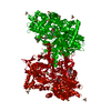
 Components Components | Glycogen phosphorylase, muscle form | ||||||
 Keywords Keywords | TRANSFERASE / Phosphorylase / Inhibitor / EGCG / Caffeine | ||||||
| Function / homology |  Function and homology information Function and homology informationglycogen phosphorylase / glycogen phosphorylase activity / glycogen catabolic process / skeletal muscle myofibril / pyridoxal phosphate binding / nucleotide binding Similarity search - Function | ||||||
| Biological species |  | ||||||
| Method |  X-RAY DIFFRACTION / SYNCHROTRON / MOLECULAR REPLACEMENT / Resolution: 2.5 Å X-RAY DIFFRACTION / SYNCHROTRON / MOLECULAR REPLACEMENT / Resolution: 2.5 Å | ||||||
 Authors Authors | Alexopoulos, S. / Papakostopoulou, S. / Koulas, M.S. / Leonidas, D.D. / Skamnaki, V. | ||||||
| Funding support | 1items
| ||||||
 Citation Citation | Journal: J.Agric.Food Chem. / Year: 2024 Title: Evidence for the Quercetin Binding Site of Glycogen Phosphorylase as a Target for Liver-Isoform-Selective Inhibitors against Glioblastoma: Investigation of Flavanols Epigallocatechin Gallate and Epigallocatechin. Authors: Alexopoulos, S. / McGawley, M. / Mathews, R. / Papakostopoulou, S. / Koulas, S. / Leonidas, D.D. / Zwain, T. / Hayes, J.M. / Skamnaki, V. | ||||||
| History |
|
- Structure visualization
| Structure viewer | Molecule: MolmilJmol/JSmol |
|---|
- Downloads & links
-Download
| PDBx/mmCIF format | 8r6v.cif.gz | 196.3 KB | Display | PDBx/mmCIF format |
|---|---|---|---|---|
| PDB format | pdb8r6v.ent.gz | 150 KB | Display | PDB format |
| PDBx/mmJSON format | 8r6v.json.gz | Tree view | PDBx/mmJSON format | |
| Others |  Other downloads Other downloads |
-Validation report
| Arichive directory | https://data.pdbj.org/pub/pdb/validation_reports/r6/8r6vftp://data.pdbj.org/pub/pdb/validation_reports/r6/8r6v | HTTPS FTP |
|---|
-Related structure data


-Links
 PDBj
PDBj
- Assembly
| Deposited unit | 
| ||||||||||||
|---|---|---|---|---|---|---|---|---|---|---|---|---|---|
| 1 | 
| ||||||||||||
| Unit cell |
| ||||||||||||
| Components on special symmetry positions |
|
-Components
| #1: Protein | Mass: 96197.836 Da / Num. of mol.: 1 / Source method: isolated from a natural source / Source: (natural) | ||||||||||
|---|---|---|---|---|---|---|---|---|---|---|---|
| #2: Chemical | ChemComp-DMS /   Mass: 78.133 Da / Num. of mol.: 29 / Source method: obtained synthetically / Formula: C2H6OS / Comment: DMSO, precipitant*YM Mass: 78.133 Da / Num. of mol.: 29 / Source method: obtained synthetically / Formula: C2H6OS / Comment: DMSO, precipitant*YM#3: Chemical | ChemComp-KDH / ( | 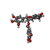  Mass: 458.372 Da / Num. of mol.: 1 / Source method: isolated from a natural source / Formula: C22H18O11 / Feature type: SUBJECT OF INVESTIGATION Mass: 458.372 Da / Num. of mol.: 1 / Source method: isolated from a natural source / Formula: C22H18O11 / Feature type: SUBJECT OF INVESTIGATION#4: Chemical | ChemComp-CFF / | 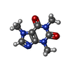  Mass: 194.191 Da / Num. of mol.: 1 / Source method: obtained synthetically / Formula: C8H10N4O2 / Comment: medication*YM Mass: 194.191 Da / Num. of mol.: 1 / Source method: obtained synthetically / Formula: C8H10N4O2 / Comment: medication*YM#5: Water | ChemComp-HOH / |  Mass: 18.015 Da / Num. of mol.: 474 / Source method: isolated from a natural source / Formula: H2O Mass: 18.015 Da / Num. of mol.: 474 / Source method: isolated from a natural source / Formula: H2OHas ligand of interest | Y | Has protein modification | Y | |
-Experimental details
-Experiment
| Experiment | Method: X-RAY DIFFRACTION / Number of used crystals: 1 |
|---|
- Sample preparation
| Crystal | Density Matthews: 2.44 Å3/Da / Density % sol: 49.61 % |
|---|---|
| Crystal grow | Temperature: 289 K / Method: batch mode / pH: 6.7 / Details: 10mM BES buffer pH 6.7 |
-Data collection
| Diffraction | Mean temperature: 100 K / Serial crystal experiment: N |
|---|---|
| Diffraction source | Source: SYNCHROTRON / Site: PETRA III, EMBL c/o DESY  / Beamline: P13 (MX1) / Wavelength: 0.9763 Å / Beamline: P13 (MX1) / Wavelength: 0.9763 Å |
| Detector | Type: DECTRIS PILATUS 6M / Detector: PIXEL / Date: Mar 14, 2022 |
| Radiation | Monochromator: M / Protocol: SINGLE WAVELENGTH / Monochromatic (M) / Laue (L): M / Scattering type: x-ray |
| Radiation wavelength | Wavelength: 0.9763 Å / Relative weight: 1 |
| Reflection | Resolution: 2.5→126.44 Å / Num. obs: 32975 / % possible obs: 100 % / Redundancy: 10.6 % / CC1/2: 0.998 / Net I/σ(I): 18.6 |
| Reflection shell | Resolution: 2.5→2.6 Å / Redundancy: 10.4 % / Mean I/σ(I) obs: 9.4 / Num. unique obs: 3649 / CC1/2: 0.986 |
- Processing
| Software |
| |||||||||||||||||||||
|---|---|---|---|---|---|---|---|---|---|---|---|---|---|---|---|---|---|---|---|---|---|---|
| Refinement | Method to determine structure: MOLECULAR REPLACEMENT / Resolution: 2.5→89.4 Å / SU B: 15.128 / SU ML: 0.184 / Cross valid method: THROUGHOUT / ESU R: 0.81 / ESU R Free: 0.288 / Details: HYDROGENS HAVE BEEN USED IF PRESENT IN THE INPUT
| |||||||||||||||||||||
| Displacement parameters | Biso mean: 25.941 Å2
| |||||||||||||||||||||
| Refinement step | Cycle: LAST / Resolution: 2.5→89.4 Å
| |||||||||||||||||||||
| LS refinement shell | Resolution: 2.5→2.565 Å
|
