 Movie
Movie Controller

[English] 日本語
 Yorodumi
- PDB-8r5t: Crystal structure of NDM-1 in complex with benzobisheterocycle co... -
+ Open data

- Basic information
| Entry | Database: PDB / ID: 8r5t | ||||||
|---|---|---|---|---|---|---|---|
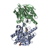
| Title | Crystal structure of NDM-1 in complex with benzobisheterocycle compound 14. | ||||||
 Components Components | Metallo-beta-lactamase type 2 | ||||||
 Keywords Keywords | ANTIMICROBIAL PROTEIN / metallo-beta-lactamase / inhibitor | ||||||
| Function / homology |  Function and homology information Function and homology informationantibiotic catabolic process / beta-lactamase activity / beta-lactamase / periplasmic space / response to antibiotic / zinc ion binding Similarity search - Function | ||||||
| Biological species |  Klebsiella pneumoniae (bacteria) Klebsiella pneumoniae (bacteria) | ||||||
| Method |  X-RAY DIFFRACTION / SYNCHROTRON / FOURIER SYNTHESIS / Resolution: 1.58 Å X-RAY DIFFRACTION / SYNCHROTRON / FOURIER SYNTHESIS / Resolution: 1.58 Å | ||||||
 Authors Authors | Hinchliffe, P. / Spencer, J. | ||||||
| Funding support |  United States, 1items United States, 1items
| ||||||
 Citation Citation | Journal: J.Med.Chem. / Year: 2024 Title: Rational Design of Benzobisheterocycle Metallo-beta-Lactamase Inhibitors: A Tricyclic Scaffold Enhances Potency against Target Enzymes. Authors: Villamil, V. / Rossi, M.A. / Mojica, M.F. / Hinchliffe, P. / Martinez, V. / Castillo, V. / Saiz, C. / Banchio, C. / Macias, M.A. / Spencer, J. / Bonomo, R.A. / Vila, A. / Moreno, D.M. / Mahler, G. | ||||||
| History |
|
- Structure visualization
| Structure viewer | Molecule: MolmilJmol/JSmol |
|---|
- Downloads & links
-Download
| PDBx/mmCIF format | 8r5t.cif.gz | 262.9 KB | Display | PDBx/mmCIF format |
|---|---|---|---|---|
| PDB format | pdb8r5t.ent.gz | 214.3 KB | Display | PDB format |
| PDBx/mmJSON format | 8r5t.json.gz | Tree view | PDBx/mmJSON format | |
| Others |  Other downloads Other downloads |
-Validation report
| Summary document | 8r5t_validation.pdf.gz | 779.3 KB | Display | wwPDB validaton report |
|---|---|---|---|---|
| Full document | 8r5t_full_validation.pdf.gz | 781.5 KB | Display | |
| Data in XML | 8r5t_validation.xml.gz | 21.7 KB | Display | |
| Data in CIF | 8r5t_validation.cif.gz | 32 KB | Display | |
| Arichive directory | https://data.pdbj.org/pub/pdb/validation_reports/r5/8r5tftp://data.pdbj.org/pub/pdb/validation_reports/r5/8r5t | HTTPS FTP |
-Related structure data
-Links
 PDBj
PDBj

- Assembly
| Deposited unit | 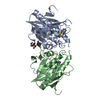
| ||||||||
|---|---|---|---|---|---|---|---|---|---|
| 1 | 
| ||||||||
| 2 | 
| ||||||||
| Unit cell |
|
-Components
| #1: Protein | Mass: 25775.951 Da / Num. of mol.: 2 Source method: isolated from a genetically manipulated source Source: (gene. exp.) Klebsiella pneumoniae (bacteria) / Gene: blaNDM-1 / Production host: #2: Chemical | ChemComp-Y4E / [( | Mass: 224.346 Da / Num. of mol.: 1 / Source method: obtained synthetically / Formula: C10H12N2S2 / Feature type: SUBJECT OF INVESTIGATION #3: Chemical | ChemComp-ZN /   Mass: 65.409 Da / Num. of mol.: 4 / Source method: obtained synthetically / Formula: Zn Mass: 65.409 Da / Num. of mol.: 4 / Source method: obtained synthetically / Formula: Zn#4: Chemical |   Mass: 96.063 Da / Num. of mol.: 2 / Source method: obtained synthetically / Formula: SO4 Mass: 96.063 Da / Num. of mol.: 2 / Source method: obtained synthetically / Formula: SO4#5: Water | ChemComp-HOH / |  Mass: 18.015 Da / Num. of mol.: 343 / Source method: isolated from a natural source / Formula: H2O Mass: 18.015 Da / Num. of mol.: 343 / Source method: isolated from a natural source / Formula: H2OHas ligand of interest | Y | |
|---|
-Experimental details
-Experiment
| Experiment | Method: X-RAY DIFFRACTION / Number of used crystals: 1 |
|---|
- Sample preparation
| Crystal | Density Matthews: 1.95 Å3/Da / Density % sol: 36.83 % |
|---|---|
| Crystal grow | Temperature: 292 K / Method: vapor diffusion, sitting drop / pH: 5.8 Details: 0.1M bis-tris pH 5.8, 32% PEG 3350, 0.15M (NH4)2SO4 |
-Data collection
| Diffraction | Mean temperature: 100 K / Serial crystal experiment: N |
|---|---|
| Diffraction source | Source: SYNCHROTRON / Site: Diamond  / Beamline: I03 / Wavelength: 0.87 Å / Beamline: I03 / Wavelength: 0.87 Å |
| Detector | Type: DECTRIS PILATUS3 6M / Detector: PIXEL / Date: Oct 22, 2019 |
| Radiation | Protocol: SINGLE WAVELENGTH / Monochromatic (M) / Laue (L): M / Scattering type: x-ray |
| Radiation wavelength | Wavelength: 0.87 Å / Relative weight: 1 |
| Reflection | Resolution: 1.58→35.05 Å / Num. obs: 55898 / % possible obs: 100 % / Redundancy: 13.6 % / CC1/2: 0.999 / Rpim(I) all: 0.032 / Net I/σ(I): 13.4 |
| Reflection shell | Resolution: 1.58→1.61 Å / Mean I/σ(I) obs: 1.6 / Num. unique obs: 2738 / CC1/2: 0.668 / Rpim(I) all: 0.586 |
- Processing
| Software |
| |||||||||||||||||||||||||||||||||||||||||||||||||||||||||||||||||||||||||||||||||||||||||||||||||||||||||
|---|---|---|---|---|---|---|---|---|---|---|---|---|---|---|---|---|---|---|---|---|---|---|---|---|---|---|---|---|---|---|---|---|---|---|---|---|---|---|---|---|---|---|---|---|---|---|---|---|---|---|---|---|---|---|---|---|---|---|---|---|---|---|---|---|---|---|---|---|---|---|---|---|---|---|---|---|---|---|---|---|---|---|---|---|---|---|---|---|---|---|---|---|---|---|---|---|---|---|---|---|---|---|---|---|---|---|
| Refinement | Method to determine structure: FOURIER SYNTHESIS / Resolution: 1.58→34.356 Å / SU ML: 0.18 / Cross valid method: THROUGHOUT / σ(F): 1.34 / Phase error: 21.1 / Stereochemistry target values: ML
| |||||||||||||||||||||||||||||||||||||||||||||||||||||||||||||||||||||||||||||||||||||||||||||||||||||||||
| Solvent computation | Shrinkage radii: 0.9 Å / VDW probe radii: 1.11 Å / Solvent model: FLAT BULK SOLVENT MODEL | |||||||||||||||||||||||||||||||||||||||||||||||||||||||||||||||||||||||||||||||||||||||||||||||||||||||||
| Displacement parameters | Biso max: 110.42 Å2 / Biso mean: 28.6065 Å2 / Biso min: 12.51 Å2 | |||||||||||||||||||||||||||||||||||||||||||||||||||||||||||||||||||||||||||||||||||||||||||||||||||||||||
| Refinement step | Cycle: final / Resolution: 1.58→34.356 Å
| |||||||||||||||||||||||||||||||||||||||||||||||||||||||||||||||||||||||||||||||||||||||||||||||||||||||||
| LS refinement shell | Refine-ID: X-RAY DIFFRACTION / Rfactor Rfree error: 0 / % reflection obs: 100 %
|
