 Movie
Movie Controller


+ Open data

- Basic information
| Entry | Database: PDB / ID: 8r1a | |||||||||
|---|---|---|---|---|---|---|---|---|---|---|
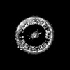
| Title | Model of the membrane-bound GBP1 oligomer | |||||||||
 Components Components | Guanylate binding protein 1 | |||||||||
 Keywords Keywords | ANTIMICROBIAL PROTEIN / Oligomer / GTPase / Interferon-induced | |||||||||
| Function / homology |  Function and homology information Function and homology information | |||||||||
| Biological species |  Homo sapiens (human) Homo sapiens (human) | |||||||||
| Method | ELECTRON MICROSCOPY / subtomogram averaging / cryo EM / Resolution: 26.8 Å | |||||||||
 Authors Authors | Weismehl, M. / Chu, X. / Kutsch, M. / Lauterjung, P. / Herrmann, C. / Kudryashev, M. / Daumke, O. | |||||||||
| Funding support |  Germany, 2items Germany, 2items
| |||||||||
 Citation Citation | Journal: EMBO J / Year: 2024 Title: Structural insights into the activation mechanism of antimicrobial GBP1. Authors: Marius Weismehl / Xiaofeng Chu / Miriam Kutsch / Paul Lauterjung / Christian Herrmann / Misha Kudryashev / Oliver Daumke /  Abstract: The dynamin-related human guanylate-binding protein 1 (GBP1) mediates host defenses against microbial pathogens. Upon GTP binding and hydrolysis, auto-inhibited GBP1 monomers dimerize and assemble ...The dynamin-related human guanylate-binding protein 1 (GBP1) mediates host defenses against microbial pathogens. Upon GTP binding and hydrolysis, auto-inhibited GBP1 monomers dimerize and assemble into soluble and membrane-bound oligomers, which are crucial for innate immune responses. How higher-order GBP1 oligomers are built from dimers, and how assembly is coordinated with nucleotide-dependent conformational changes, has remained elusive. Here, we present cryo-electron microscopy-based structural data of soluble and membrane-bound GBP1 oligomers, which show that GBP1 assembles in an outstretched dimeric conformation. We identify a surface-exposed helix in the large GTPase domain that contributes to the oligomerization interface, and we probe its nucleotide- and dimerization-dependent movements that facilitate the formation of an antimicrobial protein coat on a gram-negative bacterial pathogen. Our results reveal a sophisticated activation mechanism for GBP1, in which nucleotide-dependent structural changes coordinate dimerization, oligomerization, and membrane binding to allow encapsulation of pathogens within an antimicrobial protein coat. | |||||||||
| History |
|
- Structure visualization
| Structure viewer | Molecule: MolmilJmol/JSmol |
|---|
- Downloads & links
-Download
| PDBx/mmCIF format | 8r1a.cif.gz | 601.3 KB | Display | PDBx/mmCIF format |
|---|---|---|---|---|
| PDB format | pdb8r1a.ent.gz | 485.7 KB | Display | PDB format |
| PDBx/mmJSON format | 8r1a.json.gz | Tree view | PDBx/mmJSON format | |
| Others |  Other downloads Other downloads |
-Validation report
| Arichive directory | https://data.pdbj.org/pub/pdb/validation_reports/r1/8r1aftp://data.pdbj.org/pub/pdb/validation_reports/r1/8r1a | HTTPS FTP |
|---|
-Related structure data
| Related structure data | 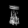 18806MC M: map data used to model this data C: citing same article ( |
|---|---|
| Similar structure data |
-Links
 PDBj
PDBj

- Assembly
| Deposited unit | 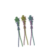
|
|---|---|
| 1 |
|
-Components
| #1: Protein | Mass: 69267.930 Da / Num. of mol.: 6 Source method: isolated from a genetically manipulated source Source: (gene. exp.) Homo sapiens (human) / Production host:  #2: Chemical | ChemComp-AF3 /   Mass: 83.977 Da / Num. of mol.: 6 / Source method: obtained synthetically / Formula: AlF3 Mass: 83.977 Da / Num. of mol.: 6 / Source method: obtained synthetically / Formula: AlF3#3: Chemical | ChemComp-MG /   Mass: 24.305 Da / Num. of mol.: 6 / Source method: obtained synthetically / Formula: Mg Mass: 24.305 Da / Num. of mol.: 6 / Source method: obtained synthetically / Formula: Mg#4: Chemical | ChemComp-GDP /   Type: RNA linking / Mass: 443.201 Da / Num. of mol.: 6 / Source method: obtained synthetically / Formula: C10H15N5O11P2 / Comment: GDP, energy-carrying molecule*YM Type: RNA linking / Mass: 443.201 Da / Num. of mol.: 6 / Source method: obtained synthetically / Formula: C10H15N5O11P2 / Comment: GDP, energy-carrying molecule*YMHas ligand of interest | N | |
|---|
-Experimental details
-Experiment
| Experiment | Method: ELECTRON MICROSCOPY |
|---|---|
| EM experiment | Aggregation state: PARTICLE / 3D reconstruction method: subtomogram averaging |
- Sample preparation
| Component | Name: Membrane-bound oligomer of human guanylate-binding protein 1 (GBP1) Type: COMPLEX / Details: in complex with GDP-AlFx / Entity ID: #1 / Source: RECOMBINANT |
|---|---|
| Source (natural) | Organism: Homo sapiens (human) |
| Source (recombinant) | Organism: |
| Buffer solution | pH: 7.9 Details: 50 mM Tris-HCl, 150 mM NaCl, 5 mM MgCl2 (supplemented with 200 uM GDP, 300 uM AlCl3, 10 mM NaF) |
| Specimen | Conc.: 0.68 mg/ml / Embedding applied: NO / Shadowing applied: NO / Staining applied: NO / Vitrification applied: YES |
| Vitrification | Cryogen name: ETHANE |
- Electron microscopy imaging
| Experimental equipment |  Model: Titan Krios / Image courtesy: FEI Company |
|---|---|
| Microscopy | Model: FEI TITAN KRIOS |
| Electron gun | Electron source:  FIELD EMISSION GUN / Accelerating voltage: 300 kV / Illumination mode: FLOOD BEAM FIELD EMISSION GUN / Accelerating voltage: 300 kV / Illumination mode: FLOOD BEAM |
| Electron lens | Mode: BRIGHT FIELD / Nominal magnification: 42000 X / Nominal defocus max: 5000 nm / Nominal defocus min: 2000 nm |
| Image recording | Electron dose: 2.8 e/Å2 / Avg electron dose per subtomogram: 130 e/Å2 / Film or detector model: GATAN K3 (6k x 4k) Details: Tilt series were acquired with a Hybrid-STA (Sanchez et al. 2020, Nat Commun): exposure dose of 2.8 e-/A2 for non-zero tilted projection and 14.4 e-/A2 for zero tilted projection |
- Processing
| CTF correction | Type: PHASE FLIPPING ONLY |
|---|---|
| Symmetry | Point symmetry: C1 (asymmetric) |
| 3D reconstruction | Resolution: 26.8 Å / Resolution method: FSC 0.143 CUT-OFF / Num. of particles: 6146 / Symmetry type: POINT |
| EM volume selection | Num. of tomograms: 104 / Num. of volumes extracted: 70160 |
