 Movie
Movie Controller

[English] 日本語
 Yorodumi
- PDB-8qyc: Zorya anti-bacteriophage defense system ZorD in complex with ATP-... -
+ Open data

- Basic information
| Entry | Database: PDB / ID: 8qyc | ||||||||||||||||||||||||||||||||||||||||||||||||||||||
|---|---|---|---|---|---|---|---|---|---|---|---|---|---|---|---|---|---|---|---|---|---|---|---|---|---|---|---|---|---|---|---|---|---|---|---|---|---|---|---|---|---|---|---|---|---|---|---|---|---|---|---|---|---|---|---|
| Title | Zorya anti-bacteriophage defense system ZorD in complex with ATP-gamma-S | ||||||||||||||||||||||||||||||||||||||||||||||||||||||
 Components Components | DEAD/DEAH box helicase | ||||||||||||||||||||||||||||||||||||||||||||||||||||||
 Keywords Keywords | ANTIVIRAL PROTEIN / Nuclease | ||||||||||||||||||||||||||||||||||||||||||||||||||||||
| Function / homology | PHOSPHOTHIOPHOSPHORIC ACID-ADENYLATE ESTER / :  Function and homology information Function and homology information | ||||||||||||||||||||||||||||||||||||||||||||||||||||||
| Biological species |  | ||||||||||||||||||||||||||||||||||||||||||||||||||||||
| Method | ELECTRON MICROSCOPY / single particle reconstruction / cryo EM / Resolution: 2.75 Å | ||||||||||||||||||||||||||||||||||||||||||||||||||||||
 Authors Authors | Hu, H. / Taylor, N.M.I. | ||||||||||||||||||||||||||||||||||||||||||||||||||||||
| Funding support |  Denmark, 3items Denmark, 3items
| ||||||||||||||||||||||||||||||||||||||||||||||||||||||
 Citation Citation | Journal: Nature / Year: 2025 Title: Structure and mechanism of the Zorya anti-phage defence system. Authors: Haidai Hu / Philipp F Popp / Thomas C D Hughes / Aritz Roa-Eguiara / Nicole R Rutbeek / Freddie J O Martin / Ivo Alexander Hendriks / Leighton J Payne / Yumeng Yan / Dorentina Humolli / ...Authors: Haidai Hu / Philipp F Popp / Thomas C D Hughes / Aritz Roa-Eguiara / Nicole R Rutbeek / Freddie J O Martin / Ivo Alexander Hendriks / Leighton J Payne / Yumeng Yan / Dorentina Humolli / Victor Klein-Sousa / Inga Songailiene / Yong Wang / Michael Lund Nielsen / Richard M Berry / Alexander Harms / Marc Erhardt / Simon A Jackson / Nicholas M I Taylor /      Abstract: Zorya is a recently identified and widely distributed bacterial immune system that protects bacteria from viral (phage) infections. Three Zorya subtypes have been identified, each containing ...Zorya is a recently identified and widely distributed bacterial immune system that protects bacteria from viral (phage) infections. Three Zorya subtypes have been identified, each containing predicted membrane-embedded ZorA-ZorB (ZorAB) complexes paired with soluble subunits that differ among Zorya subtypes, notably ZorC and ZorD in type I Zorya systems. Here we investigate the molecular basis of Zorya defence using cryo-electron microscopy, mutagenesis, fluorescence microscopy, proteomics and functional studies. We present cryo-electron microscopy structures of ZorAB and show that it shares stoichiometry and features of other 5:2 inner membrane ion-driven rotary motors. The ZorAB complex contains a dimeric ZorB peptidoglycan-binding domain and a pentameric α-helical coiled-coil tail made of ZorA that projects approximately 70 nm into the cytoplasm. We also characterize the structure and function of the soluble Zorya components ZorC and ZorD, finding that they have DNA-binding and nuclease activity, respectively. Comprehensive functional and mutational analyses demonstrate that all Zorya components work in concert to protect bacterial cells against invading phages. We provide evidence that ZorAB operates as a proton-driven motor that becomes activated after sensing of phage invasion. Subsequently, ZorAB transfers the phage invasion signal through the ZorA cytoplasmic tail to recruit and activate the soluble ZorC and ZorD effectors, which facilitate the degradation of the phage DNA. In summary, our study elucidates the foundational mechanisms of Zorya function as an anti-phage defence system. | ||||||||||||||||||||||||||||||||||||||||||||||||||||||
| History |
|
- Structure visualization
| Structure viewer | Molecule: MolmilJmol/JSmol |
|---|
- Downloads & links
-Download
| PDBx/mmCIF format | 8qyc.cif.gz | 193.1 KB | Display | PDBx/mmCIF format |
|---|---|---|---|---|
| PDB format | pdb8qyc.ent.gz | 147.7 KB | Display | PDB format |
| PDBx/mmJSON format | 8qyc.json.gz | Tree view | PDBx/mmJSON format | |
| Others |  Other downloads Other downloads |
-Validation report
| Arichive directory | https://data.pdbj.org/pub/pdb/validation_reports/qy/8qycftp://data.pdbj.org/pub/pdb/validation_reports/qy/8qyc | HTTPS FTP |
|---|
-Related structure data
| Related structure data |  18750MC 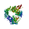 8qy7C 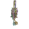 8qydC 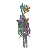 8qyhC 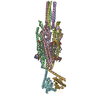 8qykC 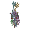 8qyyC 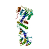 8r68C M: map data used to model this data C: citing same article ( |
|---|---|
| Similar structure data |

-Links
 PDBj
PDBj
- Assembly
| Deposited unit | 
|
|---|---|
| 1 |
|
-Components
| #1: Protein | Mass: 122880.039 Da / Num. of mol.: 1 Source method: isolated from a genetically manipulated source Source: (gene. exp.) |
|---|---|
| #2: Chemical | ChemComp-AGS /   Mass: 523.247 Da / Num. of mol.: 1 / Source method: obtained synthetically / Formula: C10H16N5O12P3S / Feature type: SUBJECT OF INVESTIGATION / Comment: ATP-gamma-S, energy-carrying molecule analogue*YM Mass: 523.247 Da / Num. of mol.: 1 / Source method: obtained synthetically / Formula: C10H16N5O12P3S / Feature type: SUBJECT OF INVESTIGATION / Comment: ATP-gamma-S, energy-carrying molecule analogue*YM |
| Has ligand of interest | Y |
| Has protein modification | N |
-Experimental details
-Experiment
| Experiment | Method: ELECTRON MICROSCOPY |
|---|---|
| EM experiment | Aggregation state: PARTICLE / 3D reconstruction method: single particle reconstruction |
- Sample preparation
| Component | Name: Zorya anti-bacteriophage defense system ZorD in complex with ATP-gamma-S Type: ORGANELLE OR CELLULAR COMPONENT / Entity ID: #1 / Source: RECOMBINANT |
|---|---|
| Source (natural) | Organism: |
| Source (recombinant) | Organism: |
| Buffer solution | pH: 7.5 |
| Specimen | Embedding applied: NO / Shadowing applied: NO / Staining applied: NO / Vitrification applied: YES |
| Vitrification | Cryogen name: ETHANE |
- Electron microscopy imaging
| Experimental equipment |  Model: Titan Krios / Image courtesy: FEI Company |
|---|---|
| Microscopy | Model: FEI TITAN KRIOS |
| Electron gun | Electron source:  FIELD EMISSION GUN / Accelerating voltage: 300 kV / Illumination mode: SPOT SCAN FIELD EMISSION GUN / Accelerating voltage: 300 kV / Illumination mode: SPOT SCAN |
| Electron lens | Mode: BRIGHT FIELD / Nominal defocus max: 2000 nm / Nominal defocus min: 500 nm |
| Image recording | Electron dose: 40 e/Å2 / Film or detector model: FEI FALCON II (4k x 4k) |
- Processing
| EM software | Name: PHENIX / Category: model refinement | ||||||||||||||||||||||||
|---|---|---|---|---|---|---|---|---|---|---|---|---|---|---|---|---|---|---|---|---|---|---|---|---|---|
| CTF correction | Type: NONE | ||||||||||||||||||||||||
| 3D reconstruction | Resolution: 2.75 Å / Resolution method: FSC 0.143 CUT-OFF / Num. of particles: 211477 / Symmetry type: POINT | ||||||||||||||||||||||||
| Refine LS restraints |
|
