[English] 日本語
Yorodumi
- EMDB-18752: EcZorAB_WT ZorB PGBDs Local refinement -

+
Open data


ID or keywords:

Loading...

-
Basic information

Entry
Database: EMDB / ID: EMD-18752
TitleEcZorAB_WT ZorB PGBDs Local refinement
Map data
Sample
  • Organelle or cellular component: Zorya anti-bacteriophage defense system ZorAB
    • Protein or peptide: ZorB PGBDs
KeywordsNuclease / ANTIVIRAL PROTEIN
Biological speciesEscherichia coli (E. coli)
Methodsingle particle reconstruction / cryo EM / Resolution: 3.51 Å
AuthorsHaidai H / Nicholas MIT
Funding support Denmark, 3 items
OrganizationGrant numberCountry
Novo Nordisk FoundationNNF17OC0031006 Denmark
Novo Nordisk FoundationNNF21OC0071948 Denmark
LundbeckfondenR347-2020-2429 Denmark
Citation
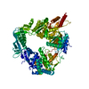
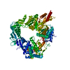
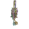
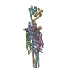
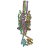
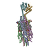
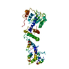
Journal: Nature / Year: 2025
Title: Structure and mechanism of the Zorya anti-phage defence system.
Authors: Haidai Hu / Philipp F Popp / Thomas C D Hughes / Aritz Roa-Eguiara / Nicole R Rutbeek / Freddie J O Martin / Ivo Alexander Hendriks / Leighton J Payne / Yumeng Yan / Dorentina Humolli / ...Authors: Haidai Hu / Philipp F Popp / Thomas C D Hughes / Aritz Roa-Eguiara / Nicole R Rutbeek / Freddie J O Martin / Ivo Alexander Hendriks / Leighton J Payne / Yumeng Yan / Dorentina Humolli / Victor Klein-Sousa / Inga Songailiene / Yong Wang / Michael Lund Nielsen / Richard M Berry / Alexander Harms / Marc Erhardt / Simon A Jackson / Nicholas M I Taylor /
Abstract: Zorya is a recently identified and widely distributed bacterial immune system that protects bacteria from viral (phage) infections. Three Zorya subtypes have been identified, each containing ...Zorya is a recently identified and widely distributed bacterial immune system that protects bacteria from viral (phage) infections. Three Zorya subtypes have been identified, each containing predicted membrane-embedded ZorA-ZorB (ZorAB) complexes paired with soluble subunits that differ among Zorya subtypes, notably ZorC and ZorD in type I Zorya systems. Here we investigate the molecular basis of Zorya defence using cryo-electron microscopy, mutagenesis, fluorescence microscopy, proteomics and functional studies. We present cryo-electron microscopy structures of ZorAB and show that it shares stoichiometry and features of other 5:2 inner membrane ion-driven rotary motors. The ZorAB complex contains a dimeric ZorB peptidoglycan-binding domain and a pentameric α-helical coiled-coil tail made of ZorA that projects approximately 70 nm into the cytoplasm. We also characterize the structure and function of the soluble Zorya components ZorC and ZorD, finding that they have DNA-binding and nuclease activity, respectively. Comprehensive functional and mutational analyses demonstrate that all Zorya components work in concert to protect bacterial cells against invading phages. We provide evidence that ZorAB operates as a proton-driven motor that becomes activated after sensing of phage invasion. Subsequently, ZorAB transfers the phage invasion signal through the ZorA cytoplasmic tail to recruit and activate the soluble ZorC and ZorD effectors, which facilitate the degradation of the phage DNA. In summary, our study elucidates the foundational mechanisms of Zorya function as an anti-phage defence system.
History
DepositionOct 25, 2023-
Header (metadata) releaseNov 6, 2024-
Map releaseNov 6, 2024-
UpdateMay 21, 2025-
Current statusMay 21, 2025Processing site: PDBe / Status: Released

-
Structure visualization

Supplemental images

Downloads & links

-
Map

FileDownload / File: emd_18752.map.gz / Format: CCP4 / Size: 476.8 MB / Type: IMAGE STORED AS FLOATING POINT NUMBER (4 BYTES)
Projections & slices

Image control

Size
Brightness
Contrast
Others
AxesZ (Sec.)Y (Row.)X (Col.)
0.83 Å/pix.
x 500 pix.
= 416. Å
0.83 Å/pix.
x 500 pix.
= 416. Å
0.83 Å/pix.
x 500 pix.
= 416. Å

Surface

Projections

Slices (1/3)

Slices (1/2)

Slices (2/3)

Images are generated by Spider.

Voxel sizeX=Y=Z: 0.832 Å
Density
Contour LevelBy AUTHOR: 0.4
Minimum - Maximum-2.394514 - 3.8731995
Average (Standard dev.)-0.000120502664 (±0.028470613)
SymmetrySpace group: 1
Details

EMDB XML:

Map geometry
Axis orderXYZ
Origin000
Dimensions500500500
Spacing500500500
CellA=B=C: 416.0 Å
α=β=γ: 90.0 °

-
Supplemental data

-
Half map: #2

Fileemd_18752_half_map_1.map
Projections & Slices
AxesZYX

Projections

Slices (1/2)
Density Histograms

-
Half map: #1

Fileemd_18752_half_map_2.map
Projections & Slices
AxesZYX

Projections

Slices (1/2)
Density Histograms

-
Sample components

-
Entire : Zorya anti-bacteriophage defense system ZorAB

EntireName: Zorya anti-bacteriophage defense system ZorAB
Components
  • Organelle or cellular component: Zorya anti-bacteriophage defense system ZorAB
    • Protein or peptide: ZorB PGBDs

-
Supramolecule #1: Zorya anti-bacteriophage defense system ZorAB

SupramoleculeName: Zorya anti-bacteriophage defense system ZorAB / type: organelle_or_cellular_component / ID: 1 / Parent: 0 / Macromolecule list: all
Source (natural)Organism: Escherichia coli (E. coli)

-
Macromolecule #1: ZorB PGBDs

MacromoleculeName: ZorB PGBDs / type: protein_or_peptide / ID: 1 / Enantiomer: LEVO
Source (natural)Organism: Escherichia coli (E. coli)
SequenceString: MFGNAFGVKK RRSDEAEKPF WISYADLMTA MMVLFLVVMV ASLSSVTQRI QRAEQGEKAR GQDISRLCER LELHARNVNK NIVVDCHDNR ISFGEAGRFA HNQFFLNAEG QKALQDVVPL VLEASNSEEG KKWFKQIVIE GFTDTDGSYL YNLHLSLQRS EWVMCSLLDS ...String:
MFGNAFGVKK RRSDEAEKPF WISYADLMTA MMVLFLVVMV ASLSSVTQRI QRAEQGEKAR GQDISRLCER LELHARNVNK NIVVDCHDNR ISFGEAGRFA HNQFFLNAEG QKALQDVVPL VLEASNSEEG KKWFKQIVIE GFTDTDGSYL YNLHLSLQRS EWVMCSLLDS RSPLQKNISA EQQLQIRKLF LAGGVSFNNA KESKEASRRV ELRMQFFGLK DKRDKADEVD FPPVVNKEVC QLVMPL

-
Experimental details

-
Structure determination

Methodcryo EM
Processingsingle particle reconstruction
Aggregation stateparticle

-
Sample preparation

BufferpH: 7.5
VitrificationCryogen name: ETHANE

-
Electron microscopy

MicroscopeFEI TITAN KRIOS
Image recordingFilm or detector model: FEI FALCON II (4k x 4k) / Average electron dose: 40.0 e/Å2
Electron beamAcceleration voltage: 300 kV / Electron source: FIELD EMISSION GUN
Electron opticsIllumination mode: SPOT SCAN / Imaging mode: BRIGHT FIELD / Nominal defocus max: 2.0 µm / Nominal defocus min: 0.5 µm
Experimental equipment
Model: Titan Krios / Image courtesy: FEI Company

+
Image processing

CTF correctionType: NONE
Startup modelType of model: NONE
Final reconstructionResolution.type: BY AUTHOR / Resolution: 3.51 Å / Resolution method: FSC 0.143 CUT-OFF / Number images used: 227728
Initial angle assignmentType: ANGULAR RECONSTITUTION
Final angle assignmentType: PROJECTION MATCHING
FSC plot (resolution estimation)

+
About Yorodumi

-
News

-
Feb 9, 2022. New format data for meta-information of EMDB entries

New format data for meta-information of EMDB entries

  • Version 3 of the EMDB header file is now the official format.
  • The previous official version 1.9 will be removed from the archive.

Related info.:EMDB header

External links:wwPDB to switch to version 3 of the EMDB data model

-
Aug 12, 2020. Covid-19 info

Covid-19 info

URL: https://pdbj.org/emnavi/covid19.php

New page: Covid-19 featured information page in EM Navigator.

Related info.:Covid-19 info / Mar 5, 2020. Novel coronavirus structure data

+
Mar 5, 2020. Novel coronavirus structure data

Novel coronavirus structure data

Related info.:Yorodumi Speices / Aug 12, 2020. Covid-19 info

External links:COVID-19 featured content - PDBj / Molecule of the Month (242):Coronavirus Proteases

+
Jan 31, 2019. EMDB accession codes are about to change! (news from PDBe EMDB page)

EMDB accession codes are about to change! (news from PDBe EMDB page)

  • The allocation of 4 digits for EMDB accession codes will soon come to an end. Whilst these codes will remain in use, new EMDB accession codes will include an additional digit and will expand incrementally as the available range of codes is exhausted. The current 4-digit format prefixed with “EMD-” (i.e. EMD-XXXX) will advance to a 5-digit format (i.e. EMD-XXXXX), and so on. It is currently estimated that the 4-digit codes will be depleted around Spring 2019, at which point the 5-digit format will come into force.
  • The EM Navigator/Yorodumi systems omit the EMD- prefix.

Related info.:Q: What is EMD? / ID/Accession-code notation in Yorodumi/EM Navigator

External links:EMDB Accession Codes are Changing Soon! / Contact to PDBj

+
Jul 12, 2017. Major update of PDB

Major update of PDB

  • wwPDB released updated PDB data conforming to the new PDBx/mmCIF dictionary.
  • This is a major update changing the version number from 4 to 5, and with Remediation, in which all the entries are updated.
  • In this update, many items about electron microscopy experimental information are reorganized (e.g. em_software).
  • Now, EM Navigator and Yorodumi are based on the updated data.

External links:wwPDB Remediation / Enriched Model Files Conforming to OneDep Data Standards Now Available in the PDB FTP Archive

-
Yorodumi

Thousand views of thousand structures

  • Yorodumi is a browser for structure data from EMDB, PDB, SASBDB, etc.
  • This page is also the successor to EM Navigator detail page, and also detail information page/front-end page for Omokage search.
  • The word "yorodu" (or yorozu) is an old Japanese word meaning "ten thousand". "mi" (miru) is to see.

Related info.:EMDB / PDB / SASBDB / Comparison of 3 databanks / Yorodumi Search / Aug 31, 2016. New EM Navigator & Yorodumi / Yorodumi Papers / Jmol/JSmol / Function and homology information / Changes in new EM Navigator and Yorodumi

Read more