 Movie
Movie Controller


+ Open data

- Basic information
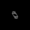
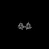
| Entry | Database: PDB / ID: 8qy5 | |||||||||||||||
|---|---|---|---|---|---|---|---|---|---|---|---|---|---|---|---|---|
| Title | Structure of interleukin 6. | |||||||||||||||
 Components Components |
| |||||||||||||||
 Keywords Keywords | IMMUNE SYSTEM / interleukin / gp130 | |||||||||||||||
| Function / homology |  Function and homology information Function and homology informationoncostatin-M receptor activity / ciliary neurotrophic factor binding / regulation of astrocyte activation / glucagon secretion / positive regulation of interleukin-21 production / IL-6-type cytokine receptor ligand interactions / MAPK3 (ERK1) activation / MAPK1 (ERK2) activation / Interleukin-27 signaling / leukemia inhibitory factor receptor activity ...oncostatin-M receptor activity / ciliary neurotrophic factor binding / regulation of astrocyte activation / glucagon secretion / positive regulation of interleukin-21 production / IL-6-type cytokine receptor ligand interactions / MAPK3 (ERK1) activation / MAPK1 (ERK2) activation / Interleukin-27 signaling / leukemia inhibitory factor receptor activity / regulation of glucagon secretion / interleukin-6 binding / hepatic immune response / Interleukin-6 signaling / negative regulation of primary miRNA processing / Interleukin-35 Signalling / regulation of vascular endothelial growth factor production / negative regulation of interleukin-1-mediated signaling pathway / triglyceride mobilization / type I oncostatin-M receptor complex / interleukin-27 receptor activity / oncostatin-M-mediated signaling pathway / regulation of microglial cell activation / ciliary neurotrophic factor receptor activity / ciliary neurotrophic factor receptor binding / leukemia inhibitory factor signaling pathway / interleukin-11 receptor activity / interleukin-11 binding / interleukin-6 receptor activity / negative regulation of interleukin-6-mediated signaling pathway / ciliary neurotrophic factor receptor complex / ciliary neurotrophic factor-mediated signaling pathway / interleukin-6 receptor complex / positive regulation of apoptotic DNA fragmentation / interleukin-27-mediated signaling pathway / hepatocyte proliferation / positive regulation of type B pancreatic cell apoptotic process / germinal center B cell differentiation / neutrophil apoptotic process / response to peptidoglycan / positive regulation of extracellular matrix disassembly / interleukin-11-mediated signaling pathway / positive regulation of B cell activation / positive regulation of receptor signaling pathway via STAT / interleukin-6 receptor binding / positive regulation of T-helper 2 cell cytokine production / regulation of Notch signaling pathway / T-helper 17 cell lineage commitment / inflammatory response to wounding / endocrine pancreas development / regulation of neuroinflammatory response / negative regulation of collagen biosynthetic process / positive regulation of acute inflammatory response / positive regulation of astrocyte differentiation / vascular endothelial growth factor production / negative regulation of chemokine production / positive regulation of adaptive immune response / T follicular helper cell differentiation / intestinal epithelial cell development / positive regulation of leukocyte chemotaxis / positive regulation of glomerular mesangial cell proliferation / negative regulation of interleukin-8 production / positive regulation of peptidyl-tyrosine phosphorylation / positive regulation of platelet aggregation / neutrophil mediated immunity / cell surface receptor signaling pathway via STAT / cytokine receptor activity / positive regulation of cytokine production involved in inflammatory response / negative regulation of bone resorption / positive regulation of leukocyte adhesion to vascular endothelial cell / CD163 mediating an anti-inflammatory response / positive regulation of smooth muscle cell migration / Interleukin-6 signaling / positive regulation of neuroinflammatory response / glycogen metabolic process / interleukin-6-mediated signaling pathway / growth factor binding / positive regulation of immunoglobulin production / positive regulation of Notch signaling pathway / MAPK3 (ERK1) activation / negative regulation of fat cell differentiation / negative regulation of cytosolic calcium ion concentration / MAPK1 (ERK2) activation / neuronal cell body membrane / cytokine binding / Interleukin-10 signaling / maintenance of blood-brain barrier / positive regulation of interleukin-17 production / regulation of insulin secretion / humoral immune response / Transcriptional Regulation by VENTX / protein tyrosine kinase activator activity / positive regulation of interleukin-10 production / monocyte chemotaxis / negative regulation of lipid storage / positive regulation of peptidyl-serine phosphorylation / regulation of angiogenesis / positive regulation of vascular endothelial growth factor production / positive regulation of glial cell proliferation / positive regulation of osteoblast differentiation Similarity search - Function | |||||||||||||||
| Biological species |   Homo sapiens (human) Homo sapiens (human) | |||||||||||||||
| Method | ELECTRON MICROSCOPY / single particle reconstruction / cryo EM / Resolution: 3.1 Å | |||||||||||||||
 Authors Authors | Gardner, S. / Bubeck, D. / Jin, Y. | |||||||||||||||
| Funding support |  United Kingdom, European Union, 4items United Kingdom, European Union, 4items
| |||||||||||||||
 Citation Citation | Journal: Nat Commun / Year: 2024 Title: Structural insights into IL-11-mediated signalling and human IL6ST variant-associated immunodeficiency. Authors: Scott Gardner / Yibo Jin / Paul K Fyfe / Tomas B Voisin / Junel Sotolongo Bellón / Elizabeth Pohler / Jacob Piehler / Ignacio Moraga / Doryen Bubeck /  Abstract: IL-11 and IL-6 activate signalling via assembly of the cell surface receptor gp130; however, it is unclear how signals are transmitted across the membrane to instruct cellular responses. Here we ...IL-11 and IL-6 activate signalling via assembly of the cell surface receptor gp130; however, it is unclear how signals are transmitted across the membrane to instruct cellular responses. Here we solve the cryoEM structure of the IL-11 receptor recognition complex to discover how differences in gp130-binding interfaces may drive signalling outcomes. We explore how mutations in the IL6ST gene encoding for gp130, which cause severe immune deficiencies in humans, impair signalling without blocking cytokine binding. We use cryoEM to solve structures of both IL-11 and IL-6 complexes with a mutant form of gp130 associated with human disease. Together with molecular dynamics simulations, we show that the disease-associated variant led to an increase in flexibility including motion within the cytokine-binding core and increased distance between extracellular domains. However, these distances are minimized as the transmembrane helix exits the membrane, suggesting a stringency in geometry for signalling and dimmer switch mode of action. #1: Journal: Acta Crystallogr D Struct Biol / Year: 2019 Title: Macromolecular structure determination using X-rays, neutrons and electrons: recent developments in Phenix. Authors: Dorothee Liebschner / Pavel V Afonine / Matthew L Baker / Gábor Bunkóczi / Vincent B Chen / Tristan I Croll / Bradley Hintze / Li Wei Hung / Swati Jain / Airlie J McCoy / Nigel W Moriarty ...Authors: Dorothee Liebschner / Pavel V Afonine / Matthew L Baker / Gábor Bunkóczi / Vincent B Chen / Tristan I Croll / Bradley Hintze / Li Wei Hung / Swati Jain / Airlie J McCoy / Nigel W Moriarty / Robert D Oeffner / Billy K Poon / Michael G Prisant / Randy J Read / Jane S Richardson / David C Richardson / Massimo D Sammito / Oleg V Sobolev / Duncan H Stockwell / Thomas C Terwilliger / Alexandre G Urzhumtsev / Lizbeth L Videau / Christopher J Williams / Paul D Adams /   Abstract: Diffraction (X-ray, neutron and electron) and electron cryo-microscopy are powerful methods to determine three-dimensional macromolecular structures, which are required to understand biological ...Diffraction (X-ray, neutron and electron) and electron cryo-microscopy are powerful methods to determine three-dimensional macromolecular structures, which are required to understand biological processes and to develop new therapeutics against diseases. The overall structure-solution workflow is similar for these techniques, but nuances exist because the properties of the reduced experimental data are different. Software tools for structure determination should therefore be tailored for each method. Phenix is a comprehensive software package for macromolecular structure determination that handles data from any of these techniques. Tasks performed with Phenix include data-quality assessment, map improvement, model building, the validation/rebuilding/refinement cycle and deposition. Each tool caters to the type of experimental data. The design of Phenix emphasizes the automation of procedures, where possible, to minimize repetitive and time-consuming manual tasks, while default parameters are chosen to encourage best practice. A graphical user interface provides access to many command-line features of Phenix and streamlines the transition between programs, project tracking and re-running of previous tasks. | |||||||||||||||
| History |
|
- Structure visualization
| Structure viewer | Molecule: MolmilJmol/JSmol |
|---|
- Downloads & links
-Download
| PDBx/mmCIF format | 8qy5.cif.gz | 901.3 KB | Display | PDBx/mmCIF format |
|---|---|---|---|---|
| PDB format | pdb8qy5.ent.gz | 596.8 KB | Display | PDB format |
| PDBx/mmJSON format | 8qy5.json.gz | Tree view | PDBx/mmJSON format | |
| Others |  Other downloads Other downloads |
-Validation report
| Arichive directory | https://data.pdbj.org/pub/pdb/validation_reports/qy/8qy5ftp://data.pdbj.org/pub/pdb/validation_reports/qy/8qy5 | HTTPS FTP |
|---|
-Related structure data
| Related structure data | 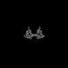 18742MC  8qy4C 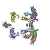 8qy6C M: map data used to model this data C: citing same article ( |
|---|---|
| Similar structure data |
-Links
 PDBj
PDBj


- Assembly
| Deposited unit | 
| ||||||||||||||||||||||||||||||||||||||||||||||||||||||||||||||||||||||||||||||||||||||||||||||||||||||||||||||||||||||||||||||||||||
|---|---|---|---|---|---|---|---|---|---|---|---|---|---|---|---|---|---|---|---|---|---|---|---|---|---|---|---|---|---|---|---|---|---|---|---|---|---|---|---|---|---|---|---|---|---|---|---|---|---|---|---|---|---|---|---|---|---|---|---|---|---|---|---|---|---|---|---|---|---|---|---|---|---|---|---|---|---|---|---|---|---|---|---|---|---|---|---|---|---|---|---|---|---|---|---|---|---|---|---|---|---|---|---|---|---|---|---|---|---|---|---|---|---|---|---|---|---|---|---|---|---|---|---|---|---|---|---|---|---|---|---|---|---|
| 1 |
| ||||||||||||||||||||||||||||||||||||||||||||||||||||||||||||||||||||||||||||||||||||||||||||||||||||||||||||||||||||||||||||||||||||
| Noncrystallographic symmetry (NCS) | NCS domain:
NCS domain segments:
|