 ムービー
ムービー コントローラー


+ データを開く

- 基本情報
基本情報
| 登録情報 | データベース: PDB / ID: 8qxb | ||||||
|---|---|---|---|---|---|---|---|
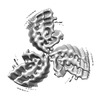
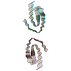
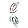
| タイトル | TDP-43 amyloid fibrils: Morphology-2 | ||||||
 要素 要素 | TAR DNA-binding protein 43 TAR DNA結合タンパク質43 TAR DNA結合タンパク質43 | ||||||
 キーワード キーワード | PROTEIN FIBRIL / Amyloidosis (アミロイドーシス) / Protein misfolding disease / Amyloid fibrils (アミロイド) / Cryo electron microscopy / Amyloid key | ||||||
| 機能・相同性 |  機能・相同性情報 機能・相同性情報nuclear inner membrane organization / interchromatin granule / perichromatin fibrils / 3'-UTR-mediated mRNA destabilization / 3'-UTR-mediated mRNA stabilization / intracellular non-membrane-bounded organelle / negative regulation by host of viral transcription / pre-mRNA intronic binding / response to endoplasmic reticulum stress / RNA splicing ...nuclear inner membrane organization / interchromatin granule / perichromatin fibrils / 3'-UTR-mediated mRNA destabilization / 3'-UTR-mediated mRNA stabilization / intracellular non-membrane-bounded organelle / negative regulation by host of viral transcription / pre-mRNA intronic binding / response to endoplasmic reticulum stress / RNA splicing / negative regulation of protein phosphorylation / molecular condensate scaffold activity / mRNA 3'-UTR binding / regulation of protein stability / regulation of circadian rhythm / positive regulation of insulin secretion / 転写後修飾 / cytoplasmic stress granule / positive regulation of protein import into nucleus / rhythmic process / 遺伝子発現の調節 / double-stranded DNA binding / regulation of apoptotic process / amyloid fibril formation / regulation of cell cycle / nuclear speck / RNA polymerase II cis-regulatory region sequence-specific DNA binding / negative regulation of gene expression / lipid binding / ミトコンドリア / DNA binding / RNA binding / 核質 / identical protein binding / 細胞核類似検索 - 分子機能 | ||||||
| 生物種 |  Homo sapiens (ヒト) Homo sapiens (ヒト) | ||||||
| 手法 | 電子顕微鏡法 / らせん対称体再構成法 / クライオ電子顕微鏡法 / 解像度: 3.86 Å | ||||||
 データ登録者 データ登録者 | Sharma, K. / Shenoy, J. / Loquet, A. / Schmidt, M. / Faendrich, M. | ||||||
| 資金援助 | 1件
| ||||||
 引用 引用 | ジャーナル: Nat Commun / 年: 2024 タイトル: Cryo-EM observation of the amyloid key structure of polymorphic TDP-43 amyloid fibrils. 著者: Kartikay Sharma / Fabian Stockert / Jayakrishna Shenoy / Mélanie Berbon / Muhammed Bilal Abdul-Shukkoor / Birgit Habenstein / Antoine Loquet / Matthias Schmidt / Marcus Fändrich /   要旨: The transactive response DNA-binding protein-43 (TDP-43) is a multi-facet protein involved in phase separation, RNA-binding, and alternative splicing. In the context of neurodegenerative diseases, ...The transactive response DNA-binding protein-43 (TDP-43) is a multi-facet protein involved in phase separation, RNA-binding, and alternative splicing. In the context of neurodegenerative diseases, abnormal aggregation of TDP-43 has been linked to amyotrophic lateral sclerosis and frontotemporal lobar degeneration through the aggregation of its C-terminal domain. Here, we report a cryo-electron microscopy (cryo-EM)-based structural characterization of TDP-43 fibrils obtained from the full-length protein. We find that the fibrils are polymorphic and contain three different amyloid structures. The structures differ in the number and relative orientation of the protofilaments, although they share a similar fold containing an amyloid key motif. The observed fibril structures differ from previously described conformations of TDP-43 fibrils and help to better understand the structural landscape of the amyloid fibril structures derived from this protein. | ||||||
| 履歴 |
|
- 構造の表示
構造の表示
| 構造ビューア | 分子: MolmilJmol/JSmol |
|---|
- ダウンロードとリンク
-ダウンロード
| PDBx/mmCIF形式 | 8qxb.cif.gz | 196.2 KB | 表示 | PDBx/mmCIF形式 |
|---|---|---|---|---|
| PDB形式 | pdb8qxb.ent.gz | 114.9 KB | 表示 | PDB形式 |
| PDBx/mmJSON形式 | 8qxb.json.gz | ツリー表示 | PDBx/mmJSON形式 | |
| その他 |  その他のダウンロード その他のダウンロード |
-検証レポート
| アーカイブディレクトリ | https://data.pdbj.org/pub/pdb/validation_reports/qx/8qxbftp://data.pdbj.org/pub/pdb/validation_reports/qx/8qxb | HTTPS FTP |
|---|
-関連構造データ


-リンク
 PDBj
PDBj- 集合体
集合体
| 登録構造単位 | 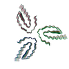
|
|---|---|
| 1 |
|
-要素
| #1: タンパク質 | TAR DNA結合タンパク質43 / TDP-43 分子量: 44784.742 Da / 分子数: 18 / 由来タイプ: 組換発現 / 由来: (組換発現) Homo sapiens (ヒト) / 遺伝子: TARDBP, TDP43 / 発現宿主:  Escherichia coli (大腸菌) / 参照: UniProt: Q13148 Escherichia coli (大腸菌) / 参照: UniProt: Q13148 |
|---|
-実験情報
-実験
| 実験 | 手法: 電子顕微鏡法 |
|---|---|
| EM実験 | 試料の集合状態: HELICAL ARRAY / 3次元再構成法: らせん対称体再構成法 |
- 試料調製
試料調製
| 構成要素 | 名称: TDP-43 amyloid fibrils: Morphology-2 / タイプ: COMPLEX / 詳細: In vitro formed TDP-43 amyloid fibrils / Entity ID: all / 由来: RECOMBINANT |
|---|---|
| 分子量 | 実験値: NO |
| 由来(天然) | 生物種: Homo sapiens (ヒト) |
| 由来(組換発現) | 生物種: Escherichia coli (大腸菌) |
| 緩衝液 | pH: 7.4 |
| 試料 | 包埋: NO / シャドウイング: NO / 染色: NO / 凍結: YES |
| 試料支持 | グリッドの材料: COPPER / グリッドのサイズ: 400 divisions/in. / グリッドのタイプ: C-flat-1.2/1.3 |
| 急速凍結 | 装置: GATAN CRYOPLUNGE 3 / 凍結剤: ETHANE / 湿度: 85 % |
- 電子顕微鏡撮影
| 実験機器 |  モデル: Titan Krios / 画像提供: FEI Company |
|---|---|
| 顕微鏡 | モデル: FEI TITAN KRIOS |
| 電子銃 | 電子線源: FIELD EMISSION GUN / 加速電圧: 300 kV / 照射モード: FLOOD BEAM |
| 電子レンズ | モード: BRIGHT FIELDBright-field microscopy / 最大 デフォーカス(公称値): 2500 nm / 最小 デフォーカス(公称値): 1000 nm / Cs: 2.7 mm |
| 試料ホルダ | 凍結剤: NITROGEN |
| 撮影 | 平均露光時間: 10 sec. / 電子線照射量: 52.08 e/Å2 / 検出モード: COUNTING フィルム・検出器のモデル: GATAN K2 QUANTUM (4k x 4k) 実像数: 1641 |
| 画像スキャン | 動画フレーム数/画像: 40 |
- 解析
解析
| EMソフトウェア |
| ||||||||||||||||||||||||||||||||
|---|---|---|---|---|---|---|---|---|---|---|---|---|---|---|---|---|---|---|---|---|---|---|---|---|---|---|---|---|---|---|---|---|---|
| CTF補正 | タイプ: PHASE FLIPPING AND AMPLITUDE CORRECTION | ||||||||||||||||||||||||||||||||
| らせん対称 | 回転角度/サブユニット: -1.3 ° / 軸方向距離/サブユニット: 4.773 Å / らせん対称軸の対称性: C3 | ||||||||||||||||||||||||||||||||
| 粒子像の選択 | 選択した粒子像数: 22126 | ||||||||||||||||||||||||||||||||
| 3次元再構成 | 解像度: 3.86 Å / 解像度の算出法: FSC 0.143 CUT-OFF / 粒子像の数: 14995 / 対称性のタイプ: HELICAL | ||||||||||||||||||||||||||||||||
| 原子モデル構築 | プロトコル: BACKBONE TRACE / 空間: REAL / Target criteria: correlation coefficient |
