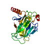
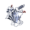
- PDB-8qwl: Structure of p53 cancer mutant Y163C -
+
データを開く
IDまたはキーワード:
読み込み中...
-
基本情報
登録情報
データベース: PDB / ID: 8qwl
タイトル
Structure of p53 cancer mutant Y163C
要素
Cellular tumor antigen p53
キーワード
DNA BINDING PROTEIN / tumor suppressor / transcription factor / cancer mutation / protein stability / conformational mutant
機能・相同性
機能・相同性情報
negative regulation of helicase activity / Loss of function of TP53 in cancer due to loss of tetramerization ability / Regulation of TP53 Expression / signal transduction by p53 class mediator / negative regulation of G1 to G0 transition / negative regulation of glucose catabolic process to lactate via pyruvate / Transcriptional activation of cell cycle inhibitor p21 / regulation of intrinsic apoptotic signaling pathway by p53 class mediator / negative regulation of pentose-phosphate shunt / ATP-dependent DNA/DNA annealing activity ...negative regulation of helicase activity / Loss of function of TP53 in cancer due to loss of tetramerization ability / Regulation of TP53 Expression / signal transduction by p53 class mediator / negative regulation of G1 to G0 transition / negative regulation of glucose catabolic process to lactate via pyruvate / Transcriptional activation of cell cycle inhibitor p21 / regulation of intrinsic apoptotic signaling pathway by p53 class mediator / negative regulation of pentose-phosphate shunt / ATP-dependent DNA/DNA annealing activity / Activation of NOXA and translocation to mitochondria / regulation of cell cycle G2/M phase transition / regulation of fibroblast apoptotic process / oligodendrocyte apoptotic process / negative regulation of miRNA processing / intrinsic apoptotic signaling pathway in response to hypoxia / positive regulation of thymocyte apoptotic process / oxidative stress-induced premature senescence / regulation of tissue remodeling / positive regulation of mitochondrial membrane permeability / mRNA transcription / bone marrow development / positive regulation of programmed necrotic cell death / circadian behavior / T cell proliferation involved in immune response / regulation of mitochondrial membrane permeability involved in apoptotic process / germ cell nucleus / RUNX3 regulates CDKN1A transcription / glucose catabolic process to lactate via pyruvate / TP53 Regulates Transcription of Death Receptors and Ligands / Activation of PUMA and translocation to mitochondria / TP53 regulates transcription of additional cell cycle genes whose exact role in the p53 pathway remain uncertain / regulation of DNA damage response, signal transduction by p53 class mediator / histone deacetylase regulator activity / negative regulation of glial cell proliferation / Regulation of TP53 Activity through Association with Co-factors / negative regulation of neuroblast proliferation / mitochondrial DNA repair / T cell lineage commitment / Formation of Senescence-Associated Heterochromatin Foci (SAHF) / ER overload response / thymocyte apoptotic process / B cell lineage commitment / TP53 Regulates Transcription of Caspase Activators and Caspases / cardiac septum morphogenesis / negative regulation of mitophagy / negative regulation of DNA replication / entrainment of circadian clock by photoperiod / negative regulation of telomere maintenance via telomerase / Zygotic genome activation (ZGA) / positive regulation of release of cytochrome c from mitochondria / PI5P Regulates TP53 Acetylation / Association of TriC/CCT with target proteins during biosynthesis / necroptotic process / TP53 Regulates Transcription of Genes Involved in Cytochrome C Release / TFIID-class transcription factor complex binding / SUMOylation of transcription factors / TP53 regulates transcription of several additional cell death genes whose specific roles in p53-dependent apoptosis remain uncertain / intrinsic apoptotic signaling pathway by p53 class mediator / negative regulation of reactive oxygen species metabolic process / rRNA transcription / Transcriptional Regulation by VENTX / cellular response to UV-C / replicative senescence / general transcription initiation factor binding / cellular response to actinomycin D / intrinsic apoptotic signaling pathway in response to endoplasmic reticulum stress / positive regulation of RNA polymerase II transcription preinitiation complex assembly / positive regulation of execution phase of apoptosis / intrinsic apoptotic signaling pathway in response to DNA damage by p53 class mediator / neuroblast proliferation / Pyroptosis / viral process / hematopoietic stem cell differentiation / response to X-ray / embryonic organ development / chromosome organization / type II interferon-mediated signaling pathway / somitogenesis / TP53 Regulates Transcription of Genes Involved in G1 Cell Cycle Arrest / hematopoietic progenitor cell differentiation / glial cell proliferation / negative regulation of fibroblast proliferation / positive regulation of cardiac muscle cell apoptotic process / core promoter sequence-specific DNA binding / negative regulation of stem cell proliferation / cellular response to glucose starvation / mitophagy / cis-regulatory region sequence-specific DNA binding / positive regulation of intrinsic apoptotic signaling pathway / Regulation of TP53 Activity through Acetylation / gastrulation / response to salt stress / 14-3-3 protein binding / mitotic G1 DNA damage checkpoint signaling / negative regulation of proteolysis / MDM2/MDM4 family protein binding / cardiac muscle cell apoptotic process / transcription repressor complex / intrinsic apoptotic signaling pathway 類似検索 - 分子機能
 ムービー
ムービー コントローラー


 データを開く
 基本情報
基本情報 要素
要素 キーワード
キーワード 機能・相同性情報
 Homo sapiens (ヒト)
 X線回折 /
  データ登録者
 ドイツ, 1件
  引用
引用 構造の表示
構造の表示 ダウンロードとリンク
 その他のダウンロード
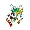
 PDBj
PDBj


 集合体
集合体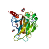


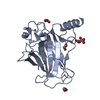
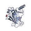
 分子量: 65.409 Da / 分子数: 1 / 由来タイプ: 合成 / 式: Zn
 分子量: 62.068 Da / 分子数: 3 / 由来タイプ: 合成 / 式: C2H6O2
 分子量: 102.046 Da / 分子数: 1 / 由来タイプ: 天然 / 式: C3H2O4
 分子量: 18.015 Da / 分子数: 198 / 由来タイプ: 天然 / 式: H2O
 試料調製
試料調製 / ビームライン: X06SA / 波長: 0.9999 Å
 解析
解析