[English] 日本語
Yorodumi
- PDB-8qvd: Deinococcus aerius TR0125 C-glucosyl deglycosidase (CGD), wild ty... -

+
Open data


ID or keywords:

Loading...

-
Basic information

Entry
Database: PDB / ID: 8qvd
TitleDeinococcus aerius TR0125 C-glucosyl deglycosidase (CGD), wild type crystal cryoprotected with glycerol
Components
  • DUF6379 domain-containing protein
  • Xylose isomerase-like TIM barrel domain-containing protein
KeywordsLYASE / bacterial C-glucosyl deglycosidase / C-C bond cleavage / C-glucosyl aromatic polyketides / C-glucosyl flavonoids / Deinococcus aerius / soil bacterium / N-terminal DUF6379 beta-sandwich domain / C-terminal TIM-barrel domain / alpha2beta2 heterotetramer
Function / homologyDomain of unknown function DUF6379 / Domain of unknown function (DUF6379) / Xylose isomerase-like, TIM barrel domain / Xylose isomerase-like TIM barrel / Xylose isomerase-like superfamily / : / Xylose isomerase-like TIM barrel domain-containing protein / DUF6379 domain-containing protein
Function and homology information
Biological speciesDeinococcus aerius (bacteria)
MethodX-RAY DIFFRACTION / SYNCHROTRON / MOLECULAR REPLACEMENT / Resolution: 3.3 Å
AuthorsFurlanetto, V. / Kalyani, D.C. / Kostelac, A. / Haltrich, D. / Hallberg, B.M. / Divne, C.
Funding support Austria, 5items
OrganizationGrant numberCountry
Other governmentFormas 2017-00983
Other privateOscar and Lili Lamm Memorial Foundation DO2017-0020
Other governmentVR 2017-03877
Austrian Science FundFWF P33545 Austria
Austrian Science FundFWF W1224 Austria
Citation
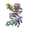
Journal: J Mol Biol / Year: 2024
Title: Structural and Functional Characterization of a Gene Cluster Responsible for Deglycosylation of C-glucosyl Flavonoids and Xanthonoids by Deinococcus aerius.
Authors: Valentina Furlanetto / Dayanand C Kalyani / Anja Kostelac / Jolanta Puc / Dietmar Haltrich / B Martin Hällberg / Christina Divne /
Abstract: Plant C-glycosylated aromatic polyketides are important for plant and animal health. These are specialized metabolites that perform functions both within the plant, and in interaction with soil or ...Plant C-glycosylated aromatic polyketides are important for plant and animal health. These are specialized metabolites that perform functions both within the plant, and in interaction with soil or intestinal microbes. Despite the importance of these plant compounds, there is still limited knowledge of how they are metabolized. The Gram-positive aerobic soil bacterium Deinococcus aerius strain TR0125 and other Deinococcus species thrive in a wide range of harsh environments. In this work, we identified a C-glycoside deglycosylation gene cluster in the genome of D. aerius. The cluster includes three genes coding for a GMC-type oxidoreductase (DaCGO1) that oxidizes the glucosyl C3 position in aromatic C-glucosyl compounds, which in turn provides the substrate for the C-glycoside deglycosidase (DaCGD; composed of α+β subunits) that cleaves the glucosyl-aglycone C-C bond. Our results from size-exclusion chromatography, single particle cryo-electron microscopy and X-ray crystallography show that DaCGD is an αβ heterotetramer, which represents a novel oligomeric state among bacterial CGDs. Importantly, the high-resolution X-ray structure of DaCGD provides valuable insights into the activation of the catalytic hydroxide ion by Lys261. DaCGO1 is specific for the 6-C-glucosyl flavones isovitexin, isoorientin and the 2-C-glucosyl xanthonoid mangiferin, and the subsequent C-C-bond cleavage by DaCGD generated apigenin, luteolin and norathyriol, respectively. Of the substrates tested, isovitexin was the preferred substrate (DaCGO1, K 0.047 mM, k 51 min; DaCGO1/DaCGD, K 0.083 mM, k 0.42 min).
History
DepositionOct 17, 2023Deposition site: PDBE / Processing site: PDBE
Revision 1.0May 29, 2024Provider: repository / Type: Initial release

-
Structure visualization

Structure viewerMolecule:
MolmilJmol/JSmol

Downloads & links

-
Assembly

Deposited unit
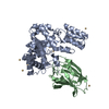
A: Xylose isomerase-like TIM barrel domain-containing protein
B: DUF6379 domain-containing protein
C: Xylose isomerase-like TIM barrel domain-containing protein
D: DUF6379 domain-containing protein
hetero molecules


Theoretical massNumber of molelcules
Total (without water)110,73519
Polymers109,0484
Non-polymers1,68615
Water00
1


  • Idetical with deposited unit
  • defined by author&software
  • Evidence: gel filtration, Oligomeric state confirmed by gel filtration, cryo-EM, crystal structure
TypeNameSymmetry operationNumber
identity operation1_555x,y,z1
Buried area9430 Å2
ΔGint-124 kcal/mol
Surface area35810 Å2
MethodPISA
Unit cell
Length a, b, c (Å)70.030, 93.780, 93.460
Angle α, β, γ (deg.)90.00, 94.94, 90.00
Int Tables number4
Space group name H-MP1211

-
Components

#1: Protein Xylose isomerase-like TIM barrel domain-containing protein


Mass: 40554.328 Da / Num. of mol.: 2
Source method: isolated from a genetically manipulated source
Source: (gene. exp.) Deinococcus aerius (bacteria) / Strain: TR0125 / Gene: DAERI_200053 / Plasmid: pNIC-CTHO / Production host: Escherichia coli B (bacteria) / Strain (production host): (DE3)-T1 / References: UniProt: A0A2I9DAN1
#2: Protein DUF6379 domain-containing protein


Mass: 13969.866 Da / Num. of mol.: 2
Source method: isolated from a genetically manipulated source
Source: (gene. exp.) Deinococcus aerius (bacteria) / Strain: TR0125 / Gene: DAERI_200052 / Plasmid: 2A-T / Production host: Escherichia coli B (bacteria) / Strain (production host): (DE3)-T1 / References: UniProt: A0A2I9E2I0
#3: Chemical
ChemComp-CD / CADMIUM ION


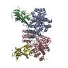
Mass: 112.411 Da / Num. of mol.: 15 / Source method: obtained synthetically / Formula: Cd
Has ligand of interestN

-
Experimental details

-
Experiment

ExperimentMethod: X-RAY DIFFRACTION / Number of used crystals: 1

-
Sample preparation

CrystalDensity Matthews: 2.96 Å3/Da / Density % sol: 58.41 %
Crystal growTemperature: 293 K / Method: vapor diffusion, sitting drop / pH: 7.5
Details: 0.1 M HEPES pH 7.5, 12% (w/v) polyethylene glycol 3,350, 5 mM CoCl2, 5 mM CdCl2, 5 mM MgCl2, and 5 mM NiCl2

-
Data collection

DiffractionMean temperature: 100 K / Serial crystal experiment: N
Diffraction sourceSource: SYNCHROTRON / Site: Diamond / Beamline: I04 / Wavelength: 0.953731 Å
DetectorType: DECTRIS EIGER2 X 16M / Detector: PIXEL / Date: Jan 27, 2023 / Details: compound refractive lenses
RadiationMonochromator: Si(111) double crystal / Protocol: SINGLE WAVELENGTH / Monochromatic (M) / Laue (L): M / Scattering type: x-ray
Radiation wavelengthWavelength: 0.953731 Å / Relative weight: 1
ReflectionResolution: 3.3→49.51 Å / Num. obs: 18261 / % possible obs: 99.8 % / Redundancy: 6.9 % / CC1/2: 0.997 / Rmerge(I) obs: 0.193 / Net I/σ(I): 7.4
Reflection shellResolution: 3.3→3.4 Å / Redundancy: 7.1 % / Mean I/σ(I) obs: 1.2 / Num. unique obs: 1557 / CC1/2: 0.637 / % possible all: 99.6

-
Processing

Software
NameVersionClassification
PHENIX(1.19.2_4158: ???)refinement
XDSdata reduction
XDSdata scaling
PHENIXphasing
RefinementMethod to determine structure: MOLECULAR REPLACEMENT / Resolution: 3.3→49.51 Å / SU ML: 0.51 / Cross valid method: FREE R-VALUE / σ(F): 1.36 / Phase error: 39.59 / Stereochemistry target values: ML
RfactorNum. reflection% reflection
Rfree0.2965 1827 10.06 %
Rwork0.2387 --
obs0.2443 18156 99.23 %
Solvent computationShrinkage radii: 0.9 Å / VDW probe radii: 1.11 Å / Solvent model: FLAT BULK SOLVENT MODEL
Refinement stepCycle: LAST / Resolution: 3.3→49.51 Å
ProteinNucleic acidLigandSolventTotal
Num. atoms7252 0 15 0 7267
Refine LS restraints
Refine-IDTypeDev idealNumber
X-RAY DIFFRACTIONf_bond_d0.0117422
X-RAY DIFFRACTIONf_angle_d1.43410024
X-RAY DIFFRACTIONf_dihedral_angle_d5.9111000
X-RAY DIFFRACTIONf_chiral_restr0.0791064
X-RAY DIFFRACTIONf_plane_restr0.0161320
LS refinement shell
Resolution (Å)Rfactor RfreeNum. reflection RfreeRfactor RworkNum. reflection RworkRefine-ID% reflection obs (%)
3.3-3.390.4511230.47841272X-RAY DIFFRACTION99
3.39-3.490.50251450.43951274X-RAY DIFFRACTION100
3.49-3.60.4331470.39741226X-RAY DIFFRACTION99
3.6-3.730.42761370.35231240X-RAY DIFFRACTION99
3.73-3.880.35541460.30521233X-RAY DIFFRACTION99
3.88-4.060.32651420.25831241X-RAY DIFFRACTION99
4.06-4.270.2811390.2311259X-RAY DIFFRACTION99
4.27-4.540.25361380.2011235X-RAY DIFFRACTION99
4.54-4.890.20451480.17081255X-RAY DIFFRACTION99
4.89-5.380.27431280.19191262X-RAY DIFFRACTION100
5.38-6.150.2711320.21941266X-RAY DIFFRACTION99
6.16-7.750.27791490.20991282X-RAY DIFFRACTION100
7.75-49.510.24851530.17961284X-RAY DIFFRACTION99
Refinement TLS params.Method: refined / Origin x: 14.0587 Å / Origin y: 16.9542 Å / Origin z: -22.1771 Å
111213212223313233
T1.4795 Å20.0142 Å20.0347 Å2-1.1563 Å20.0232 Å2--1.1473 Å2
L0.0976 °20.0277 °2-0.0229 °2-1.3117 °2-0.124 °2--1.0548 °2
S-0.0389 Å °0.0332 Å °-0.0284 Å °-0.2309 Å °-0.0765 Å °-0.0457 Å °0.0042 Å °0.2087 Å °0.1234 Å °
Refinement TLS groupSelection details: all

+
About Yorodumi

-
News

-
Feb 9, 2022. New format data for meta-information of EMDB entries

New format data for meta-information of EMDB entries

  • Version 3 of the EMDB header file is now the official format.
  • The previous official version 1.9 will be removed from the archive.

Related info.:EMDB header

External links:wwPDB to switch to version 3 of the EMDB data model

-
Aug 12, 2020. Covid-19 info

Covid-19 info

URL: https://pdbjlvh1.pdbj.org/emnavi/covid19.php

New page: Covid-19 featured information page in EM Navigator.

Related info.:Covid-19 info / Mar 5, 2020. Novel coronavirus structure data

+
Mar 5, 2020. Novel coronavirus structure data

Novel coronavirus structure data

Related info.:Yorodumi Speices / Aug 12, 2020. Covid-19 info

External links:COVID-19 featured content - PDBj / Molecule of the Month (242):Coronavirus Proteases

+
Jan 31, 2019. EMDB accession codes are about to change! (news from PDBe EMDB page)

EMDB accession codes are about to change! (news from PDBe EMDB page)

  • The allocation of 4 digits for EMDB accession codes will soon come to an end. Whilst these codes will remain in use, new EMDB accession codes will include an additional digit and will expand incrementally as the available range of codes is exhausted. The current 4-digit format prefixed with “EMD-” (i.e. EMD-XXXX) will advance to a 5-digit format (i.e. EMD-XXXXX), and so on. It is currently estimated that the 4-digit codes will be depleted around Spring 2019, at which point the 5-digit format will come into force.
  • The EM Navigator/Yorodumi systems omit the EMD- prefix.

Related info.:Q: What is EMD? / ID/Accession-code notation in Yorodumi/EM Navigator

External links:EMDB Accession Codes are Changing Soon! / Contact to PDBj

+
Jul 12, 2017. Major update of PDB

Major update of PDB

  • wwPDB released updated PDB data conforming to the new PDBx/mmCIF dictionary.
  • This is a major update changing the version number from 4 to 5, and with Remediation, in which all the entries are updated.
  • In this update, many items about electron microscopy experimental information are reorganized (e.g. em_software).
  • Now, EM Navigator and Yorodumi are based on the updated data.

External links:wwPDB Remediation / Enriched Model Files Conforming to OneDep Data Standards Now Available in the PDB FTP Archive

-
Yorodumi

Thousand views of thousand structures

  • Yorodumi is a browser for structure data from EMDB, PDB, SASBDB, etc.
  • This page is also the successor to EM Navigator detail page, and also detail information page/front-end page for Omokage search.
  • The word "yorodu" (or yorozu) is an old Japanese word meaning "ten thousand". "mi" (miru) is to see.

Related info.:EMDB / PDB / SASBDB / Comparison of 3 databanks / Yorodumi Search / Aug 31, 2016. New EM Navigator & Yorodumi / Yorodumi Papers / Jmol/JSmol / Function and homology information / Changes in new EM Navigator and Yorodumi

Read more