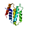
ジャーナル: Nat Struct Mol Biol / 年: 2024 タイトル: Structure of the native γ-tubulin ring complex capping spindle microtubules. 著者: Tom Dendooven / Stanislau Yatskevich / Alister Burt / Zhuo A Chen / Dom Bellini / Juri Rappsilber / John V Kilmartin / David Barford / 要旨: Microtubule (MT) filaments, composed of α/β-tubulin dimers, are fundamental to cellular architecture, function and organismal development. They are nucleated from MT organizing centers by the ...Microtubule (MT) filaments, composed of α/β-tubulin dimers, are fundamental to cellular architecture, function and organismal development. They are nucleated from MT organizing centers by the evolutionarily conserved γ-tubulin ring complex (γTuRC). However, the molecular mechanism of nucleation remains elusive. Here we used cryo-electron tomography to determine the structure of the native γTuRC capping the minus end of a MT in the context of enriched budding yeast spindles. In our structure, γTuRC presents a ring of γ-tubulin subunits to seed nucleation of exclusively 13-protofilament MTs, adopting an active closed conformation to function as a perfect geometric template for MT nucleation. Our cryo-electron tomography reconstruction revealed that a coiled-coil protein staples the first row of α/β-tubulin of the MT to alternating positions along the γ-tubulin ring of γTuRC. This positioning of α/β-tubulin onto γTuRC suggests a role for the coiled-coil protein in augmenting γTuRC-mediated MT nucleation. Based on our results, we describe a molecular model for budding yeast γTuRC activation and MT nucleation.
 ムービー
ムービー コントローラー


 データを開く
 基本情報
基本情報 要素
要素 キーワード
キーワード 機能・相同性情報
 データ登録者
 英国,
英国,  ドイツ, 3件
  引用
引用
 構造の表示
構造の表示 ダウンロードとリンク
 その他のダウンロード


 PDBj
PDBj


 集合体
集合体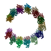
 試料調製
試料調製 電子顕微鏡撮影
 FIELD EMISSION GUN / 加速電圧: 300 kV / 照射モード: SPOT SCAN
 解析
解析