 Movie
Movie Controller

[English] 日本語
 Yorodumi
- PDB-8qtz: Cryo-EM reconstruction of VP5*/VP8* assembly from SA11 Rotavirus ... -
+ Open data

- Basic information
| Entry | Database: PDB / ID: 8qtz | ||||||
|---|---|---|---|---|---|---|---|
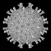
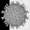
| Title | Cryo-EM reconstruction of VP5*/VP8* assembly from SA11 Rotavirus Tripsinized Triple Layered Particle | ||||||
 Components Components | Outer capsid protein VP4 | ||||||
 Keywords Keywords | VIRUS / Rotavirus / dsRNA virus | ||||||
| Function / homology |  Function and homology information Function and homology informationhost cell rough endoplasmic reticulum / permeabilization of host organelle membrane involved in viral entry into host cell / host cytoskeleton / viral outer capsid / host cell endoplasmic reticulum-Golgi intermediate compartment / virion attachment to host cell / host cell plasma membrane Similarity search - Function | ||||||
| Biological species |  Rotavirus Rotavirus | ||||||
| Method | ELECTRON MICROSCOPY / single particle reconstruction / cryo EM / Resolution: 4.27 Å | ||||||
 Authors Authors | Asensio-Cob, D. / Perez-Mata, C. / Gomez-Blanco, J. / Vargas, J. / Rodriguez, J.M. / Luque, D. | ||||||
| Funding support |  Spain, 1items Spain, 1items
| ||||||
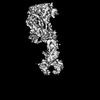
 Citation Citation | Journal: PLoS Pathog / Year: 2025 Title: Structural determinants of rotavirus proteolytic activation. Authors: Dunia Asensio-Cob / Carlos P Mata / Josue Gomez-Blanco / Javier Vargas / Javier M Rodriguez / Daniel Luque /   Abstract: The infectivity of rotavirus (RV), the leading cause of childhood diarrhea, hinges on the activation of viral particles through the proteolysis of the spike protein by trypsin-like proteases in the ...The infectivity of rotavirus (RV), the leading cause of childhood diarrhea, hinges on the activation of viral particles through the proteolysis of the spike protein by trypsin-like proteases in the host intestinal lumen. In order to determine the structural basis of trypsin activation, we have used cryogenic electron microscopy (cryo-EM) and advanced image processing methods to compare uncleaved and cleaved RV particles. We find that the conformation of the non-proteolyzed spike is constrained by the position of loops that surround its structure, linking the lectin domains of the spike head to its body. The proteolysis of these loops removes this structural constraint, thereby enabling the spike to undergo the necessary conformational changes required for cell membrane penetration. Thus, these loops function as regulatory elements to ensure that the spike protein is activated precisely when and where it is needed to facilitate a successful infection. #1: Journal: bioRxiv / Year: 2025 Title: Structural determinants of rotavirus proteolytic activation. Authors: Dunia Asensio-Cob / Carlos P Mata / Josué Gómez-Blanco / Javier Vargas / Javier M Rodríguez / Daniel Luque / Abstract: The infectivity of rotavirus (RV), the leading cause of childhood diarrhea, hinges on the activation of viral particles through the proteolysis of the spike protein by trypsin-like proteases in the ...The infectivity of rotavirus (RV), the leading cause of childhood diarrhea, hinges on the activation of viral particles through the proteolysis of the spike protein by trypsin-like proteases in the host intestinal lumen. Despite comprehensive structural characterization of the virus particle, the structural rationale behind the necessity of trypsin digestion of the VP4 protein for infectivity remains poorly understood. In this study, using cryo-electron microscopy (cryo-EM) and advanced image processing techniques, we compared uncleaved and cleaved RV virions and found that the conformation of the non-proteolyzed spike is constrained by the position of loops that surround its structure, linking the lectin domains of the spike head to its body. The proteolysis of these loops removes this structural constraint, thereby enabling the spike to undergo the necessary conformational changes required for cell membrane penetration. Thus, these loops function as regulatory elements to ensure that the spike protein is activated precisely when and where it is needed to facilitate a successful infection. | ||||||
| History |
|
- Structure visualization
| Structure viewer | Molecule: MolmilJmol/JSmol |
|---|
- Downloads & links
-Download
| PDBx/mmCIF format | 8qtz.cif.gz | 358.9 KB | Display | PDBx/mmCIF format |
|---|---|---|---|---|
| PDB format | pdb8qtz.ent.gz | Display | PDB format | |
| PDBx/mmJSON format | 8qtz.json.gz | Tree view | PDBx/mmJSON format | |
| Others |  Other downloads Other downloads |
-Validation report
| Arichive directory | https://data.pdbj.org/pub/pdb/validation_reports/qt/8qtzftp://data.pdbj.org/pub/pdb/validation_reports/qt/8qtz | HTTPS FTP |
|---|
-Related structure data
| Related structure data | 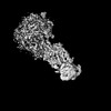 18655MC 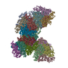 8olbC 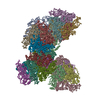 8olcC 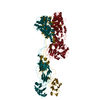 8oleC M: map data used to model this data C: citing same article ( |
|---|---|
| Similar structure data |
-Links
 PDBj
PDBj- Assembly
| Deposited unit | 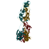
|
|---|---|
| 1 |
|
-Components
| #1: Protein | Mass: 86749.891 Da / Num. of mol.: 3 Source method: isolated from a genetically manipulated source Details: Outer capsid protein VP4 / Source: (gene. exp.) Rotavirus / Gene: VP4 / Production host:  Chlorocebus aethiops (grivet monkey) / References: UniProt: A0A060IEP4 Chlorocebus aethiops (grivet monkey) / References: UniProt: A0A060IEP4Has protein modification | N | |
|---|
-Experimental details
-Experiment
| Experiment | Method: ELECTRON MICROSCOPY |
|---|---|
| EM experiment | Aggregation state: PARTICLE / 3D reconstruction method: single particle reconstruction |
- Sample preparation
| Component | Name: Rotavirus A / Type: VIRUS / Details: Rotavirus SA11 Non-tripsinized spike / Entity ID: all / Source: NATURAL |
|---|---|
| Molecular weight | Value: 92 MDa / Experimental value: NO |
| Source (natural) | Organism: Rotavirus A / Strain: SA11 |
| Details of virus | Empty: NO / Enveloped: NO / Isolate: STRAIN / Type: VIRION |
| Natural host | Organism: Chlorocebus aethiops |
| Virus shell | Name: TLP / Diameter: 700 nm |
| Buffer solution | pH: 7.5 |
| Specimen | Embedding applied: NO / Shadowing applied: NO / Staining applied: NO / Vitrification applied: YES / Details: Rotavirus SA11 Tripsinized spike |
| Vitrification | Instrument: LEICA EM GP / Cryogen name: ETHANE / Humidity: 95 % / Chamber temperature: 295 K |
- Electron microscopy imaging
| Experimental equipment |  Model: Titan Krios / Image courtesy: FEI Company |
|---|---|
| Microscopy | Model: TFS KRIOS |
| Electron gun | Electron source:  FIELD EMISSION GUN / Accelerating voltage: 300 kV / Illumination mode: FLOOD BEAM FIELD EMISSION GUN / Accelerating voltage: 300 kV / Illumination mode: FLOOD BEAM |
| Electron lens | Mode: BRIGHT FIELD / Nominal magnification: 58000 X / Nominal defocus max: 3000 nm / Nominal defocus min: 750 nm / Alignment procedure: COMA FREE |
| Specimen holder | Cryogen: NITROGEN / Specimen holder model: FEI TITAN KRIOS AUTOGRID HOLDER |
| Image recording | Electron dose: 39.9 e/Å2 / Detector mode: INTEGRATING / Film or detector model: FEI FALCON III (4k x 4k) / Num. of real images: 2368 |
- Processing
| EM software |
| ||||||||||||||||||||||||
|---|---|---|---|---|---|---|---|---|---|---|---|---|---|---|---|---|---|---|---|---|---|---|---|---|---|
| CTF correction | Type: PHASE FLIPPING AND AMPLITUDE CORRECTION | ||||||||||||||||||||||||
| Particle selection | Num. of particles selected: 28427 | ||||||||||||||||||||||||
| 3D reconstruction | Resolution: 4.27 Å / Resolution method: FSC 0.143 CUT-OFF / Num. of particles: 22394 / Symmetry type: POINT |
