 Movie
Movie Controller

[English] 日本語
 Yorodumi
- PDB-8qmv: L2 forming RubisCO derived from ancestral sequence reconstruction... -
+ Open data

- Basic information
| Entry | Database: PDB / ID: 8qmv | |||||||||
|---|---|---|---|---|---|---|---|---|---|---|
| Title | L2 forming RubisCO derived from ancestral sequence reconstruction of the last common ancestor of Form I'' and Form I RubisCOs | |||||||||
 Components Components | RubisCO large subunit | |||||||||
 Keywords Keywords | LYASE / RubisCO / CABP / ancestral | |||||||||
| Function / homology | 2-CARBOXYARABINITOL-1,5-DIPHOSPHATE Function and homology information Function and homology information | |||||||||
| Biological species | synthetic construct (others) | |||||||||
| Method |  X-RAY DIFFRACTION / SYNCHROTRON / MOLECULAR REPLACEMENT / Resolution: 1.85 Å X-RAY DIFFRACTION / SYNCHROTRON / MOLECULAR REPLACEMENT / Resolution: 1.85 Å | |||||||||
 Authors Authors | Zarzycki, J. / Schulz, L. / Erb, T.J. / Hochberg, G.K.A. | |||||||||
| Funding support |  Germany, 2items Germany, 2items
| |||||||||
 Citation Citation | Journal: Embo J. / Year: 2025 Title: Layered entrenchment maintains essentiality in the evolution of Form I Rubisco complexes. Authors: Schulz, L. / Zarzycki, J. / Steinchen, W. / Hochberg, G.K.A. / Erb, T.J. #1: Journal: Biorxiv / Year: 2024Title: Layered entrenchment maintains essentiality in the evolution of Form I Rubisco complexes Authors: Schulz, L. / Zarzycki, J. / Steinchen, W. / Hochberg, G.K.A. / Erb, T.J. | |||||||||
| History |
|
- Structure visualization
| Structure viewer | Molecule: MolmilJmol/JSmol |
|---|
- Downloads & links
-Download
| PDBx/mmCIF format | 8qmv.cif.gz | 728.7 KB | Display | PDBx/mmCIF format |
|---|---|---|---|---|
| PDB format | pdb8qmv.ent.gz | 606.4 KB | Display | PDB format |
| PDBx/mmJSON format | 8qmv.json.gz | Tree view | PDBx/mmJSON format | |
| Others |  Other downloads Other downloads |
-Validation report
| Arichive directory | https://data.pdbj.org/pub/pdb/validation_reports/qm/8qmvftp://data.pdbj.org/pub/pdb/validation_reports/qm/8qmv | HTTPS FTP |
|---|
-Related structure data

-Links
 PDBj
PDBj
- Assembly
| Deposited unit | 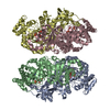
| ||||||||||||||||||
|---|---|---|---|---|---|---|---|---|---|---|---|---|---|---|---|---|---|---|---|
| 1 | 
| ||||||||||||||||||
| 2 | 
| ||||||||||||||||||
| Unit cell |
| ||||||||||||||||||
| Components on special symmetry positions |
|
-Components
| #1: Protein | Mass: 51155.840 Da / Num. of mol.: 4 Source method: isolated from a genetically manipulated source Source: (gene. exp.) synthetic construct (others) / Production host:  #2: Sugar | ChemComp-CAP /   Type: saccharide / Mass: 356.115 Da / Num. of mol.: 4 / Source method: isolated from a natural source / Formula: C6H14O13P2 / Feature type: SUBJECT OF INVESTIGATION Type: saccharide / Mass: 356.115 Da / Num. of mol.: 4 / Source method: isolated from a natural source / Formula: C6H14O13P2 / Feature type: SUBJECT OF INVESTIGATION#3: Chemical | ChemComp-MG /   Mass: 24.305 Da / Num. of mol.: 4 / Source method: obtained synthetically / Formula: Mg Mass: 24.305 Da / Num. of mol.: 4 / Source method: obtained synthetically / Formula: Mg#4: Water | ChemComp-HOH / |  Mass: 18.015 Da / Num. of mol.: 1153 / Source method: isolated from a natural source / Formula: H2O Mass: 18.015 Da / Num. of mol.: 1153 / Source method: isolated from a natural source / Formula: H2OHas ligand of interest | Y | Has protein modification | Y | |
|---|
-Experimental details
-Experiment
| Experiment | Method: X-RAY DIFFRACTION / Number of used crystals: 1 |
|---|
- Sample preparation
| Crystal | Density Matthews: 2.24 Å3/Da / Density % sol: 45 % |
|---|---|
| Crystal grow | Temperature: 289 K / Method: vapor diffusion, sitting drop / pH: 8.5 Details: Purified enzyme (10 mg/mL) in 25 mM Tricine-NaOH, 75 mM NaCl, pH 8.0 was incubated at 3% (v/v) CO2 in air and 30 degrees C for 1 hour in the presence of 0.3 mM CABP and 4.8 mM MgCl2. The ...Details: Purified enzyme (10 mg/mL) in 25 mM Tricine-NaOH, 75 mM NaCl, pH 8.0 was incubated at 3% (v/v) CO2 in air and 30 degrees C for 1 hour in the presence of 0.3 mM CABP and 4.8 mM MgCl2. The enzyme was then mixed in a 1:1 ratio with 0.1 M TRIS pH 8.5, 0.2 M magnesium chloride, 30 % (v/v) polyethylene glycol 400. The crystals were flash frozen in liquid nitrogen. |
-Data collection
| Diffraction | Mean temperature: 100 K / Serial crystal experiment: N | ||||||||||||||||||||||||||||||||||||||||||||||||||||||||||||||||||||||||||||||||||||||||||||||||||||||||||||||||||||||||||||||
|---|---|---|---|---|---|---|---|---|---|---|---|---|---|---|---|---|---|---|---|---|---|---|---|---|---|---|---|---|---|---|---|---|---|---|---|---|---|---|---|---|---|---|---|---|---|---|---|---|---|---|---|---|---|---|---|---|---|---|---|---|---|---|---|---|---|---|---|---|---|---|---|---|---|---|---|---|---|---|---|---|---|---|---|---|---|---|---|---|---|---|---|---|---|---|---|---|---|---|---|---|---|---|---|---|---|---|---|---|---|---|---|---|---|---|---|---|---|---|---|---|---|---|---|---|---|---|---|
| Diffraction source | Source: SYNCHROTRON / Site: PETRA III, EMBL c/o DESY / Beamline: P14 (MX2) / Wavelength: 0.6888 Å | ||||||||||||||||||||||||||||||||||||||||||||||||||||||||||||||||||||||||||||||||||||||||||||||||||||||||||||||||||||||||||||||
| Detector | Type: DECTRIS EIGER2 X CdTe 16M / Detector: PIXEL / Date: Dec 2, 2022 | ||||||||||||||||||||||||||||||||||||||||||||||||||||||||||||||||||||||||||||||||||||||||||||||||||||||||||||||||||||||||||||||
| Radiation | Protocol: SINGLE WAVELENGTH / Monochromatic (M) / Laue (L): M / Scattering type: x-ray | ||||||||||||||||||||||||||||||||||||||||||||||||||||||||||||||||||||||||||||||||||||||||||||||||||||||||||||||||||||||||||||||
| Radiation wavelength | Wavelength: 0.6888 Å / Relative weight: 1 | ||||||||||||||||||||||||||||||||||||||||||||||||||||||||||||||||||||||||||||||||||||||||||||||||||||||||||||||||||||||||||||||
| Reflection | Resolution: 1.85→24.97 Å / Num. obs: 155284 / % possible obs: 99.9 % / Redundancy: 13.9 % / CC1/2: 0.999 / Rmerge(I) obs: 0.113 / Rrim(I) all: 0.117 / Net I/σ(I): 16.04 | ||||||||||||||||||||||||||||||||||||||||||||||||||||||||||||||||||||||||||||||||||||||||||||||||||||||||||||||||||||||||||||||
| Reflection shell |
|
- Processing
| Software |
| |||||||||||||||||||||||||||||||||||||||||||||||||||||||||||||||||||||||||||||||||||||||||||||||||||||||||
|---|---|---|---|---|---|---|---|---|---|---|---|---|---|---|---|---|---|---|---|---|---|---|---|---|---|---|---|---|---|---|---|---|---|---|---|---|---|---|---|---|---|---|---|---|---|---|---|---|---|---|---|---|---|---|---|---|---|---|---|---|---|---|---|---|---|---|---|---|---|---|---|---|---|---|---|---|---|---|---|---|---|---|---|---|---|---|---|---|---|---|---|---|---|---|---|---|---|---|---|---|---|---|---|---|---|---|
| Refinement | Method to determine structure: MOLECULAR REPLACEMENT / Resolution: 1.85→24.97 Å / SU ML: 0.17 / Cross valid method: FREE R-VALUE / σ(F): 1.36 / Phase error: 17.98 / Stereochemistry target values: ML
| |||||||||||||||||||||||||||||||||||||||||||||||||||||||||||||||||||||||||||||||||||||||||||||||||||||||||
| Solvent computation | Shrinkage radii: 0.9 Å / VDW probe radii: 1.1 Å / Solvent model: FLAT BULK SOLVENT MODEL | |||||||||||||||||||||||||||||||||||||||||||||||||||||||||||||||||||||||||||||||||||||||||||||||||||||||||
| Refinement step | Cycle: LAST / Resolution: 1.85→24.97 Å
| |||||||||||||||||||||||||||||||||||||||||||||||||||||||||||||||||||||||||||||||||||||||||||||||||||||||||
| Refine LS restraints |
| |||||||||||||||||||||||||||||||||||||||||||||||||||||||||||||||||||||||||||||||||||||||||||||||||||||||||
| LS refinement shell |
| |||||||||||||||||||||||||||||||||||||||||||||||||||||||||||||||||||||||||||||||||||||||||||||||||||||||||
| Refinement TLS params. | Method: refined / Origin x: 17.8701 Å / Origin y: 58.0125 Å / Origin z: -19.673 Å
| |||||||||||||||||||||||||||||||||||||||||||||||||||||||||||||||||||||||||||||||||||||||||||||||||||||||||
| Refinement TLS group | Selection details: all |
