 Movie
Movie Controller

[English] 日本語
 Yorodumi
- PDB-8qjq: SmNuc1 nuclease from Stenotrophomonas maltophilia in complex with... -
+ Open data

- Basic information
| Entry | Database: PDB / ID: 8qjq | |||||||||||||||
|---|---|---|---|---|---|---|---|---|---|---|---|---|---|---|---|---|
| Title | SmNuc1 nuclease from Stenotrophomonas maltophilia in complex with cytidine - 5' - monophosphate as an inhibitor. | |||||||||||||||
 Components Components | S1/P1 Nuclease | |||||||||||||||
 Keywords Keywords | HYDROLASE / Nuclease | |||||||||||||||
| Function / homology | CYTIDINE-5'-MONOPHOSPHATE / DI(HYDROXYETHYL)ETHER / PHOSPHATE ION / :  Function and homology information Function and homology information | |||||||||||||||
| Biological species |  Stenotrophomonas maltophilia (bacteria) Stenotrophomonas maltophilia (bacteria) | |||||||||||||||
| Method |  X-RAY DIFFRACTION / SYNCHROTRON / MOLECULAR REPLACEMENT / Resolution: 1.8 Å X-RAY DIFFRACTION / SYNCHROTRON / MOLECULAR REPLACEMENT / Resolution: 1.8 Å | |||||||||||||||
 Authors Authors | Adamkova, K. / Koval, T. / Kolenko, P. / Dohnalek, J. | |||||||||||||||
| Funding support |  Czech Republic, European Union, 4items Czech Republic, European Union, 4items
| |||||||||||||||
 Citation Citation | Journal: Febs J. / Year: 2025 Title: Substrate preference, RNA binding and active site versatility of Stenotrophomonas maltophilia nuclease SmNuc1, explained by a structural study. Authors: Adamkova, K. / Trundova, M. / Koval, T. / Hustakova, B. / Kolenko, P. / Duskova, J. / Skalova, T. / Dohnalek, J. | |||||||||||||||
| History |
|
- Structure visualization
| Structure viewer | Molecule: MolmilJmol/JSmol |
|---|
- Downloads & links
-Download
| PDBx/mmCIF format | 8qjq.cif.gz | 135.7 KB | Display | PDBx/mmCIF format |
|---|---|---|---|---|
| PDB format | pdb8qjq.ent.gz | 98.7 KB | Display | PDB format |
| PDBx/mmJSON format | 8qjq.json.gz | Tree view | PDBx/mmJSON format | |
| Others |  Other downloads Other downloads |
-Validation report
| Arichive directory | https://data.pdbj.org/pub/pdb/validation_reports/qj/8qjqftp://data.pdbj.org/pub/pdb/validation_reports/qj/8qjq | HTTPS FTP |
|---|
-Related structure data
| Related structure data | 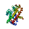 8qjlC 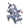 8qjmC 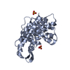 8qjnC 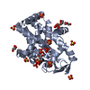 8qjoC 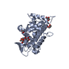 8qjpC  9emgC C: citing same article ( |
|---|---|
| Similar structure data |
-Links
 PDBj
PDBj- Assembly
| Deposited unit | 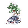
| ||||||||
|---|---|---|---|---|---|---|---|---|---|
| 1 | 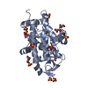
| ||||||||
| 2 | 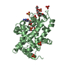
| ||||||||
| Unit cell |
|
-Components
-Protein , 1 types, 2 molecules AB
| #1: Protein | Mass: 28354.814 Da / Num. of mol.: 2 Source method: isolated from a genetically manipulated source Details: Sequence type 5, study in PMID: 28894437 Source: (gene. exp.) Stenotrophomonas maltophilia (bacteria)Strain: SanG_2012 / Gene: A1OC_03585 / Plasmid: pET303/CT-His_SmNuc1 / Details (production host): MalE signal peptide / Production host: |
|---|
-Non-polymers , 8 types, 451 molecules 


| #2: Chemical | ChemComp-ZN /  Mass: 65.409 Da / Num. of mol.: 6 / Source method: obtained synthetically / Formula: Zn / Feature type: SUBJECT OF INVESTIGATION Mass: 65.409 Da / Num. of mol.: 6 / Source method: obtained synthetically / Formula: Zn / Feature type: SUBJECT OF INVESTIGATION#3: Chemical |  Mass: 323.197 Da / Num. of mol.: 2 / Source method: obtained synthetically / Formula: C9H14N3O8P / Feature type: SUBJECT OF INVESTIGATION Mass: 323.197 Da / Num. of mol.: 2 / Source method: obtained synthetically / Formula: C9H14N3O8P / Feature type: SUBJECT OF INVESTIGATION#4: Chemical |  Mass: 92.094 Da / Num. of mol.: 2 / Source method: obtained synthetically / Formula: C3H8O3 Mass: 92.094 Da / Num. of mol.: 2 / Source method: obtained synthetically / Formula: C3H8O3#5: Chemical | ChemComp-PEG / |  Mass: 106.120 Da / Num. of mol.: 1 / Source method: obtained synthetically / Formula: C4H10O3 Mass: 106.120 Da / Num. of mol.: 1 / Source method: obtained synthetically / Formula: C4H10O3#6: Chemical | ChemComp-SO4 /  Mass: 96.063 Da / Num. of mol.: 20 / Source method: obtained synthetically / Formula: SO4 Mass: 96.063 Da / Num. of mol.: 20 / Source method: obtained synthetically / Formula: SO4#7: Chemical | ChemComp-1PE / |  Mass: 238.278 Da / Num. of mol.: 1 / Source method: obtained synthetically / Formula: C10H22O6 / Comment: precipitant*YM Mass: 238.278 Da / Num. of mol.: 1 / Source method: obtained synthetically / Formula: C10H22O6 / Comment: precipitant*YM#8: Chemical | ChemComp-PO4 / |  Mass: 94.971 Da / Num. of mol.: 1 / Source method: obtained synthetically / Formula: PO4 / Feature type: SUBJECT OF INVESTIGATION Mass: 94.971 Da / Num. of mol.: 1 / Source method: obtained synthetically / Formula: PO4 / Feature type: SUBJECT OF INVESTIGATION#9: Water | ChemComp-HOH / | Mass: 18.015 Da / Num. of mol.: 418 / Source method: isolated from a natural source / Formula: H2O |
|---|
-Details
| Has ligand of interest | Y |
|---|---|
| Has protein modification | Y |
-Experimental details
-Experiment
| Experiment | Method: X-RAY DIFFRACTION / Number of used crystals: 1 |
|---|
- Sample preparation
| Crystal | Density Matthews: 2.23 Å3/Da / Density % sol: 44.85 % |
|---|---|
| Crystal grow | Temperature: 291 K / Method: vapor diffusion, hanging drop / pH: 5.5 Details: 0.1 M Bis-Tris pH 5.5, 25 % w/v PEG 3350, 0.2 M Lithium sulfate, cryo-protection 25% v/v glycerol, protein concentration 7.5 mg/ml. Temp details: Controlled |
-Data collection
| Diffraction | Mean temperature: 100 K / Serial crystal experiment: N |
|---|---|
| Diffraction source | Source: SYNCHROTRON / Site: BESSY  / Beamline: 14.1 / Wavelength: 0.9184 Å / Beamline: 14.1 / Wavelength: 0.9184 Å |
| Detector | Type: DECTRIS PILATUS 6M / Detector: PIXEL / Date: Nov 20, 2020 |
| Radiation | Protocol: SINGLE WAVELENGTH / Monochromatic (M) / Laue (L): M / Scattering type: x-ray |
| Radiation wavelength | Wavelength: 0.9184 Å / Relative weight: 1 |
| Reflection | Resolution: 1.8→41.78 Å / Num. obs: 45358 / % possible obs: 99.4 % / Redundancy: 6.7 % / Biso Wilson estimate: 19.6 Å2 / CC1/2: 0.995 / Rmerge(I) obs: 0.108 / Rrim(I) all: 0.117 / Net I/σ(I): 14.2 |
| Reflection shell | Resolution: 1.8→1.84 Å / Redundancy: 6.4 % / Rmerge(I) obs: 1.059 / Mean I/σ(I) obs: 1.9 / Num. unique obs: 2614 / CC1/2: 0.699 / Rrim(I) all: 1.153 / % possible all: 98.9 |
- Processing
| Software |
| ||||||||||||||||||||||||||||||||||||||||||||||||||||||||||||||||||||||||||||||||||||||||||||||||||||||||||||||||||||||||||||||||||||||||||||||||||||||||||||||||||||||||||
|---|---|---|---|---|---|---|---|---|---|---|---|---|---|---|---|---|---|---|---|---|---|---|---|---|---|---|---|---|---|---|---|---|---|---|---|---|---|---|---|---|---|---|---|---|---|---|---|---|---|---|---|---|---|---|---|---|---|---|---|---|---|---|---|---|---|---|---|---|---|---|---|---|---|---|---|---|---|---|---|---|---|---|---|---|---|---|---|---|---|---|---|---|---|---|---|---|---|---|---|---|---|---|---|---|---|---|---|---|---|---|---|---|---|---|---|---|---|---|---|---|---|---|---|---|---|---|---|---|---|---|---|---|---|---|---|---|---|---|---|---|---|---|---|---|---|---|---|---|---|---|---|---|---|---|---|---|---|---|---|---|---|---|---|---|---|---|---|---|---|---|---|
| Refinement | Method to determine structure: MOLECULAR REPLACEMENT / Resolution: 1.8→41.78 Å / Cor.coef. Fo:Fc: 0.959 / SU B: 2.464 / SU ML: 0.072 / Cross valid method: FREE R-VALUE / ESU R: 0.124 Details: Hydrogens have been added in their riding positions. Last refinement cycle was performed against all reflections.
| ||||||||||||||||||||||||||||||||||||||||||||||||||||||||||||||||||||||||||||||||||||||||||||||||||||||||||||||||||||||||||||||||||||||||||||||||||||||||||||||||||||||||||
| Solvent computation | Ion probe radii: 0.8 Å / Shrinkage radii: 0.8 Å / VDW probe radii: 1.2 Å / Solvent model: MASK BULK SOLVENT | ||||||||||||||||||||||||||||||||||||||||||||||||||||||||||||||||||||||||||||||||||||||||||||||||||||||||||||||||||||||||||||||||||||||||||||||||||||||||||||||||||||||||||
| Displacement parameters | Biso mean: 29.076 Å2
| ||||||||||||||||||||||||||||||||||||||||||||||||||||||||||||||||||||||||||||||||||||||||||||||||||||||||||||||||||||||||||||||||||||||||||||||||||||||||||||||||||||||||||
| Refinement step | Cycle: LAST / Resolution: 1.8→41.78 Å
| ||||||||||||||||||||||||||||||||||||||||||||||||||||||||||||||||||||||||||||||||||||||||||||||||||||||||||||||||||||||||||||||||||||||||||||||||||||||||||||||||||||||||||
| Refine LS restraints |
| ||||||||||||||||||||||||||||||||||||||||||||||||||||||||||||||||||||||||||||||||||||||||||||||||||||||||||||||||||||||||||||||||||||||||||||||||||||||||||||||||||||||||||
| LS refinement shell |
|
