 Movie
Movie Controller

[English] 日本語
 Yorodumi
- PDB-8qj7: Cryo-EM structure of human DNA polymerase alpha-primase in pre-in... -
+ Open data

- Basic information
| Entry | Database: PDB / ID: 8qj7 | ||||||
|---|---|---|---|---|---|---|---|
| Title | Cryo-EM structure of human DNA polymerase alpha-primase in pre-initiation stage 1 | ||||||
 Components Components |
| ||||||
 Keywords Keywords | DNA BINDING PROTEIN / DNA polymerase / complex | ||||||
| Function / homology |  Function and homology information Function and homology informationribonucleotide binding / DNA primase AEP / DNA replication initiation / DNA/RNA hybrid binding / Inhibition of replication initiation of damaged DNA by RB1/E2F1 / alpha DNA polymerase:primase complex / regulation of type I interferon production / Telomere C-strand synthesis initiation / Polymerase switching / Processive synthesis on the lagging strand ...ribonucleotide binding / DNA primase AEP / DNA replication initiation / DNA/RNA hybrid binding / Inhibition of replication initiation of damaged DNA by RB1/E2F1 / alpha DNA polymerase:primase complex / regulation of type I interferon production / Telomere C-strand synthesis initiation / Polymerase switching / Processive synthesis on the lagging strand / Removal of the Flap Intermediate / lagging strand elongation / DNA replication, synthesis of primer / mitotic DNA replication initiation / Polymerase switching on the C-strand of the telomere / DNA strand elongation involved in DNA replication / leading strand elongation / G1/S-Specific Transcription / DNA synthesis involved in DNA repair / DNA replication origin binding / Activation of the pre-replicative complex / DNA replication initiation / Defective pyroptosis / double-strand break repair via nonhomologous end joining / nuclear matrix / protein import into nucleus / DNA-directed RNA polymerase activity / nuclear envelope / single-stranded DNA binding / 4 iron, 4 sulfur cluster binding / DNA-directed DNA polymerase / DNA-directed DNA polymerase activity / DNA replication / ciliary basal body / nucleotide binding / DNA repair / chromatin binding / protein kinase binding / chromatin / nucleolus / magnesium ion binding / DNA binding / zinc ion binding / nucleoplasm / membrane / metal ion binding / nucleus / cytosol Similarity search - Function | ||||||
| Biological species |  Homo sapiens (human) Homo sapiens (human) | ||||||
| Method | ELECTRON MICROSCOPY / single particle reconstruction / cryo EM / Resolution: 3.07 Å | ||||||
 Authors Authors | Yin, Z. / Pellegrini, L. | ||||||
| Funding support |  United Kingdom, 1items United Kingdom, 1items
| ||||||
 Citation Citation | Journal: FEBS J / Year: 2024 Title: CryoEM insights into RNA primer synthesis by the human primosome. Authors: Zhan Yin / Mairi L Kilkenny / De-Sheng Ker / Luca Pellegrini / Abstract: Eukaryotic DNA replication depends on the primosome - a complex of DNA polymerase alpha (Pol α) and primase - to initiate DNA synthesis by polymerisation of an RNA-DNA primer. Primer synthesis ...Eukaryotic DNA replication depends on the primosome - a complex of DNA polymerase alpha (Pol α) and primase - to initiate DNA synthesis by polymerisation of an RNA-DNA primer. Primer synthesis requires the tight coordination of primase and polymerase activities. Recent cryo-electron microscopy (cryoEM) analyses have elucidated the extensive conformational transitions required for RNA primer handover between primase and Pol α and primer elongation by Pol α. Because of the intrinsic flexibility of the primosome, however, structural information about the initiation of RNA primer synthesis is still lacking. Here, we capture cryoEM snapshots of the priming reaction to reveal the conformational trajectory of the human primosome that brings DNA primase subunits 1 and 2 (PRIM1 and PRIM2, respectively) together, poised for RNA synthesis. Furthermore, we provide experimental evidence for the continuous association of primase subunit PRIM2 with the RNA primer during primer synthesis, and for how both initiation and termination of RNA primer polymerisation are licenced by specific rearrangements of DNA polymerase alpha catalytic subunit (POLA1), the polymerase subunit of Pol α. Our findings fill a critical gap in our understanding of the conformational changes that underpin the synthesis of the RNA primer by the primosome. Together with existing evidence, they provide a complete description of the structural dynamics of the human primosome during DNA replication initiation. | ||||||
| History |
|
- Structure visualization
| Structure viewer | Molecule: MolmilJmol/JSmol |
|---|
- Downloads & links
-Download
| PDBx/mmCIF format | 8qj7.cif.gz | 915.3 KB | Display | PDBx/mmCIF format |
|---|---|---|---|---|
| PDB format | pdb8qj7.ent.gz | 750.2 KB | Display | PDB format |
| PDBx/mmJSON format | 8qj7.json.gz | Tree view | PDBx/mmJSON format | |
| Others |  Other downloads Other downloads |
-Validation report
| Arichive directory | https://data.pdbj.org/pub/pdb/validation_reports/qj/8qj7ftp://data.pdbj.org/pub/pdb/validation_reports/qj/8qj7 | HTTPS FTP |
|---|
-Related structure data
| Related structure data |  17795MC M: map data used to model this data C: citing same article ( |
|---|---|
| Similar structure data |


-Links
 PDBj
PDBj


- Assembly
| Deposited unit | 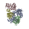
|
|---|---|
| 1 |
|
-Components
-Protein , 2 types, 2 molecules CD
| #1: Protein | Mass: 49981.012 Da / Num. of mol.: 1 Source method: isolated from a genetically manipulated source Source: (gene. exp.) Homo sapiens (human) / Gene: PRIM1 / Production host:   Spodoptera frugiperda (fall armyworm) / References: UniProt: P49642 Spodoptera frugiperda (fall armyworm) / References: UniProt: P49642 |
|---|---|
| #4: Protein | Mass: 58890.918 Da / Num. of mol.: 1 Source method: isolated from a genetically manipulated source Source: (gene. exp.) Homo sapiens (human) / Gene: PRIM2, PRIM2A / Production host: Spodoptera frugiperda (fall armyworm) / References: UniProt: P49643 |
-DNA polymerase alpha ... , 2 types, 2 molecules AB
| #2: Protein | Mass: 166131.094 Da / Num. of mol.: 1 Source method: isolated from a genetically manipulated source Source: (gene. exp.) Homo sapiens (human) / Gene: POLA1 / Production host: Spodoptera frugiperda (fall armyworm) / References: UniProt: P09884 |
|---|---|
| #3: Protein | Mass: 66015.539 Da / Num. of mol.: 1 Source method: isolated from a genetically manipulated source Source: (gene. exp.) Homo sapiens (human) / Gene: POLA2 / Production host: Spodoptera frugiperda (fall armyworm) / References: UniProt: Q14181 |
-Non-polymers , 4 types, 7 molecules 


| #5: Chemical |  Mass: 65.409 Da / Num. of mol.: 3 / Source method: obtained synthetically / Formula: Zn / Feature type: SUBJECT OF INVESTIGATION Mass: 65.409 Da / Num. of mol.: 3 / Source method: obtained synthetically / Formula: Zn / Feature type: SUBJECT OF INVESTIGATION#6: Chemical |  Mass: 54.938 Da / Num. of mol.: 2 / Source method: obtained synthetically / Formula: Mn / Feature type: SUBJECT OF INVESTIGATION Mass: 54.938 Da / Num. of mol.: 2 / Source method: obtained synthetically / Formula: Mn / Feature type: SUBJECT OF INVESTIGATION#7: Chemical | ChemComp-ATP / |  Mass: 507.181 Da / Num. of mol.: 1 / Source method: obtained synthetically / Formula: C10H16N5O13P3 / Feature type: SUBJECT OF INVESTIGATION / Comment: ATP, energy-carrying molecule*YM Mass: 507.181 Da / Num. of mol.: 1 / Source method: obtained synthetically / Formula: C10H16N5O13P3 / Feature type: SUBJECT OF INVESTIGATION / Comment: ATP, energy-carrying molecule*YM#8: Chemical | ChemComp-SF4 / |  Mass: 351.640 Da / Num. of mol.: 1 / Source method: obtained synthetically / Formula: Fe4S4 / Feature type: SUBJECT OF INVESTIGATION Mass: 351.640 Da / Num. of mol.: 1 / Source method: obtained synthetically / Formula: Fe4S4 / Feature type: SUBJECT OF INVESTIGATION |
|---|
-Details
| Has ligand of interest | Y |
|---|---|
| Has protein modification | N |
-Experimental details
-Experiment
| Experiment | Method: ELECTRON MICROSCOPY |
|---|---|
| EM experiment | Aggregation state: PARTICLE / 3D reconstruction method: single particle reconstruction |
- Sample preparation
| Component | Name: Tetrameric complex of DNA polymerase alpha-primase bound with nucleotide Type: COMPLEX / Entity ID: #1-#4 / Source: RECOMBINANT |
|---|---|
| Molecular weight | Value: 0.2875 MDa / Experimental value: NO |
| Source (natural) | Organism: Homo sapiens (human) |
| Source (recombinant) | Organism: Spodoptera frugiperda (fall armyworm) |
| Buffer solution | pH: 7.4 |
| Specimen | Conc.: 0.29 mg/ml / Embedding applied: NO / Shadowing applied: NO / Staining applied: NO / Vitrification applied: YES |
| Vitrification | Cryogen name: ETHANE |
- Electron microscopy imaging
| Experimental equipment |  Model: Titan Krios / Image courtesy: FEI Company |
|---|---|
| Microscopy | Model: FEI TITAN KRIOS |
| Electron gun | Electron source:  FIELD EMISSION GUN / Accelerating voltage: 300 kV / Illumination mode: FLOOD BEAM FIELD EMISSION GUN / Accelerating voltage: 300 kV / Illumination mode: FLOOD BEAM |
| Electron lens | Mode: BRIGHT FIELD / Nominal defocus max: 2600 nm / Nominal defocus min: 1000 nm |
| Image recording | Electron dose: 47.78 e/Å2 / Film or detector model: GATAN K3 BIOQUANTUM (6k x 4k) |
- Processing
| EM software | Name: PHENIX / Category: model refinement | ||||||||||||||||||||||||
|---|---|---|---|---|---|---|---|---|---|---|---|---|---|---|---|---|---|---|---|---|---|---|---|---|---|
| CTF correction | Type: PHASE FLIPPING AND AMPLITUDE CORRECTION | ||||||||||||||||||||||||
| 3D reconstruction | Resolution: 3.07 Å / Resolution method: FSC 0.143 CUT-OFF / Num. of particles: 1850009 / Symmetry type: POINT | ||||||||||||||||||||||||
| Refine LS restraints |
|
