- EMDB-17795: Cryo-EM structure of human DNA polymerase alpha-primase in pre-in... -
+
Open data
ID or keywords:
Loading...
-
Basic information
Entry
Database: EMDB / ID: EMD-17795
Title
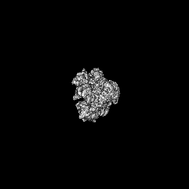
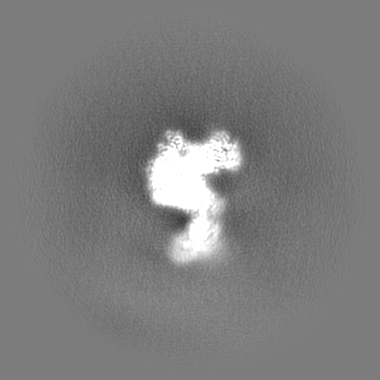
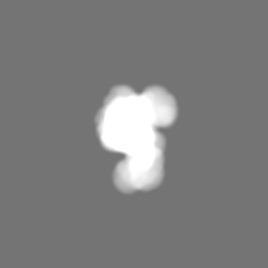
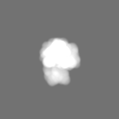
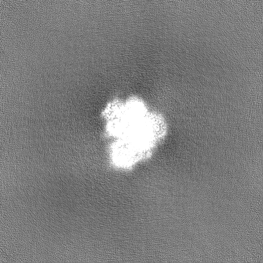
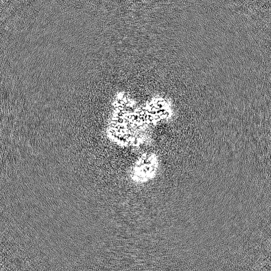
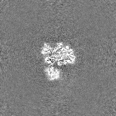
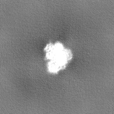
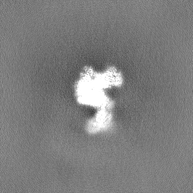
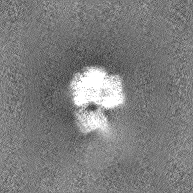
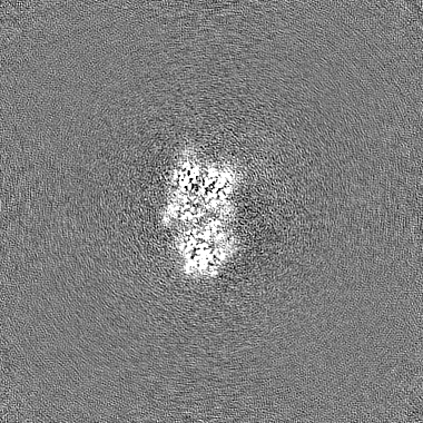
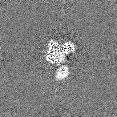
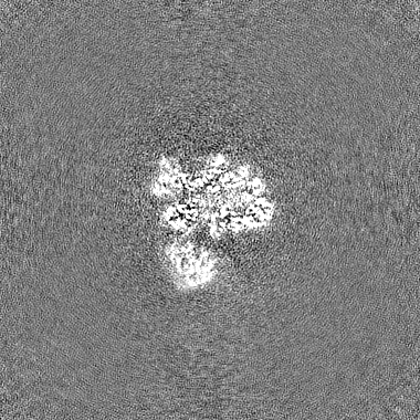
Cryo-EM structure of human DNA polymerase alpha-primase in pre-initiation stage 1
Map data
Sample
Complex: DNA polymerase alpha-primase with bound nucleotide
Keywords
DNA polymerase / complex / DNA BINDING PROTEIN
Function / homology
Function and homology information
ribonucleotide binding / DNA primase AEP / DNA replication initiation / DNA/RNA hybrid binding / Inhibition of replication initiation of damaged DNA by RB1/E2F1 / Telomere C-strand synthesis initiation / alpha DNA polymerase:primase complex / regulation of type I interferon production / Polymerase switching / Processive synthesis on the lagging strand ...ribonucleotide binding / DNA primase AEP / DNA replication initiation / DNA/RNA hybrid binding / Inhibition of replication initiation of damaged DNA by RB1/E2F1 / Telomere C-strand synthesis initiation / alpha DNA polymerase:primase complex / regulation of type I interferon production / Polymerase switching / Processive synthesis on the lagging strand / Removal of the Flap Intermediate / lagging strand elongation / DNA replication, synthesis of primer / mitotic DNA replication initiation / Polymerase switching on the C-strand of the telomere / DNA strand elongation involved in DNA replication / G1/S-Specific Transcription / leading strand elongation / DNA synthesis involved in DNA repair / DNA replication origin binding / Activation of the pre-replicative complex / DNA replication initiation / Defective pyroptosis / double-strand break repair via nonhomologous end joining / nuclear matrix / protein import into nucleus / DNA-directed RNA polymerase activity / nuclear envelope / single-stranded DNA binding / 4 iron, 4 sulfur cluster binding / DNA-directed DNA polymerase / DNA-directed DNA polymerase activity / DNA replication / ciliary basal body / nucleotide binding / DNA repair / chromatin binding / protein kinase binding / chromatin / nucleolus / magnesium ion binding / DNA binding / zinc ion binding / nucleoplasm / membrane / metal ion binding / nucleus / cytosol Similarity search - Function
DNA polymerase alpha, subunit B, N-terminal domain superfamily / : / DNA polymerase alpha subunit B, OB domain / DNA polymerase alpha subunit B N-terminal / DNA polymerase alpha, subunit B, N-terminal / DNA polymerase alpha, subunit B / : / Eukaryotic and archaeal DNA primase, large subunit N-terminal domain / DNA primase, small subunit, eukaryotic/archaeal / DNA primase large subunit, eukaryotic/archaeal ...DNA polymerase alpha, subunit B, N-terminal domain superfamily / : / DNA polymerase alpha subunit B, OB domain / DNA polymerase alpha subunit B N-terminal / DNA polymerase alpha, subunit B, N-terminal / DNA polymerase alpha, subunit B / : / Eukaryotic and archaeal DNA primase, large subunit N-terminal domain / DNA primase, small subunit, eukaryotic/archaeal / DNA primase large subunit, eukaryotic/archaeal / DNA primase, large subunit, eukaryotic / DNA primase, small subunit / DNA primase small subunit / DNA polymerase alpha catalytic subunit, N-terminal domain / DNA polymerase alpha, zinc finger domain superfamily / Eukaryotic and archaeal DNA primase, large subunit C-terminal domain / DNA Polymerase alpha zinc finger / DNA polymerase alpha subunit p180 N terminal / Zinc finger, DNA-directed DNA polymerase, family B, alpha / DNA polymerase alpha catalytic subunit, catalytic domain / DNA polymerase alpha/delta/epsilon, subunit B / DNA polymerase alpha/epsilon subunit B / DNA polymerase family B, thumb domain / DNA-directed DNA polymerase, family B, multifunctional domain / DNA-directed DNA polymerase, family B, conserved site / DNA polymerase family B signature. / DNA polymerase family B / DNA polymerase family B, exonuclease domain / DNA-directed DNA polymerase, family B, exonuclease domain / DNA polymerase, palm domain superfamily / DNA polymerase type-B family / DNA-directed DNA polymerase, family B / Ribonuclease H superfamily / Ribonuclease H-like superfamily / DNA/RNA polymerase superfamily Similarity search - Domain/homology
DNA polymerase alpha catalytic subunit / DNA primase small subunit / DNA primase large subunit / DNA polymerase alpha subunit B Similarity search - Component
Biological species
Homo sapiens (human)
Method
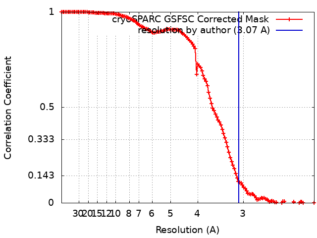
single particle reconstruction / cryo EM / Resolution: 3.07 Å
Journal: FEBS J / Year: 2024 Title: CryoEM insights into RNA primer synthesis by the human primosome. Authors: Zhan Yin / Mairi L Kilkenny / De-Sheng Ker / Luca Pellegrini / Abstract: Eukaryotic DNA replication depends on the primosome - a complex of DNA polymerase alpha (Pol α) and primase - to initiate DNA synthesis by polymerisation of an RNA-DNA primer. Primer synthesis ...Eukaryotic DNA replication depends on the primosome - a complex of DNA polymerase alpha (Pol α) and primase - to initiate DNA synthesis by polymerisation of an RNA-DNA primer. Primer synthesis requires the tight coordination of primase and polymerase activities. Recent cryo-electron microscopy (cryoEM) analyses have elucidated the extensive conformational transitions required for RNA primer handover between primase and Pol α and primer elongation by Pol α. Because of the intrinsic flexibility of the primosome, however, structural information about the initiation of RNA primer synthesis is still lacking. Here, we capture cryoEM snapshots of the priming reaction to reveal the conformational trajectory of the human primosome that brings DNA primase subunits 1 and 2 (PRIM1 and PRIM2, respectively) together, poised for RNA synthesis. Furthermore, we provide experimental evidence for the continuous association of primase subunit PRIM2 with the RNA primer during primer synthesis, and for how both initiation and termination of RNA primer polymerisation are licenced by specific rearrangements of DNA polymerase alpha catalytic subunit (POLA1), the polymerase subunit of Pol α. Our findings fill a critical gap in our understanding of the conformational changes that underpin the synthesis of the RNA primer by the primosome. Together with existing evidence, they provide a complete description of the structural dynamics of the human primosome during DNA replication initiation.
In the structure databanks used in Yorodumi, some data are registered as the other names, "COVID-19 virus" and "2019-nCoV". Here are the details of the virus and the list of structure data.
Jan 31, 2019. EMDB accession codes are about to change! (news from PDBe EMDB page)
EMDB accession codes are about to change! (news from PDBe EMDB page)
The allocation of 4 digits for EMDB accession codes will soon come to an end. Whilst these codes will remain in use, new EMDB accession codes will include an additional digit and will expand incrementally as the available range of codes is exhausted. The current 4-digit format prefixed with “EMD-” (i.e. EMD-XXXX) will advance to a 5-digit format (i.e. EMD-XXXXX), and so on. It is currently estimated that the 4-digit codes will be depleted around Spring 2019, at which point the 5-digit format will come into force.
The EM Navigator/Yorodumi systems omit the EMD- prefix.
Related info.:Q: What is EMD? / ID/Accession-code notation in Yorodumi/EM Navigator
Yorodumi is a browser for structure data from EMDB, PDB, SASBDB, etc.
This page is also the successor to EM Navigator detail page, and also detail information page/front-end page for Omokage search.
The word "yorodu" (or yorozu) is an old Japanese word meaning "ten thousand". "mi" (miru) is to see.
Related info.:EMDB / PDB / SASBDB / Comparison of 3 databanks / Yorodumi Search / Aug 31, 2016. New EM Navigator & Yorodumi / Yorodumi Papers / Jmol/JSmol / Function and homology information / Changes in new EM Navigator and Yorodumi
 Movie
Movie Controller

 Yorodumi
 Open data
 Basic information
 Map data
 Sample
 Keywords
 Function and homology information
 Homo sapiens (human)
 Authors
 United Kingdom, 1 items
  Citation
 Structure visualization
 Downloads & links
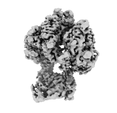 emd_17795.png
 http://ftp.pdbj.org/pub/emdb/structures/EMD-17795


 Z (Sec.)
 Y (Row.)
 X (Col.)


 Sample components
 Processing
 Electron microscopy
 FIELD EMISSION GUN