[English] 日本語
Yorodumi
- PDB-8qdv: Structure of 14-3-3 zeta delta C with the bivalent tau-pS214-pS32... -

+
Open data


ID or keywords:

Loading...

-
Basic information

Entry
Database: PDB / ID: 8qdv
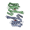
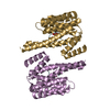
TitleStructure of 14-3-3 zeta delta C with the bivalent tau-pS214-pS324 peptide
Components
  • 14-3-3 protein zeta/delta
  • Microtubule-associated protein tau
KeywordsPROTEIN BINDING / 14-3-3 zeta / tau / protein-protein interaction
Function / homology
Function and homology information


synaptic target recognition / Golgi reassembly / NOTCH4 Activation and Transmission of Signal to the Nucleus / respiratory system process / establishment of Golgi localization / regulation of synapse maturation / tube formation / Rap1 signalling / negative regulation of protein localization to nucleus / plus-end-directed organelle transport along microtubule ...synaptic target recognition / Golgi reassembly / NOTCH4 Activation and Transmission of Signal to the Nucleus / respiratory system process / establishment of Golgi localization / regulation of synapse maturation / tube formation / Rap1 signalling / negative regulation of protein localization to nucleus / plus-end-directed organelle transport along microtubule / histone-dependent DNA binding / neurofibrillary tangle assembly / positive regulation of diacylglycerol kinase activity / axonal transport / negative regulation of establishment of protein localization to mitochondrion / neurofibrillary tangle / KSRP (KHSRP) binds and destabilizes mRNA / positive regulation of protein localization to synapse / microtubule lateral binding / tubulin complex / phosphatidylinositol bisphosphate binding / main axon / GP1b-IX-V activation signalling / regulation of long-term synaptic depression / negative regulation of kinase activity / negative regulation of tubulin deacetylation / generation of neurons / rRNA metabolic process / internal protein amino acid acetylation / regulation of chromosome organization / regulation of mitochondrial fission / axonal transport of mitochondrion / axon development / intracellular distribution of mitochondria / central nervous system neuron development / regulation of microtubule polymerization / microtubule polymerization / lipoprotein particle binding / minor groove of adenine-thymine-rich DNA binding / dynactin binding / glial cell projection / negative regulation of mitochondrial membrane potential / apolipoprotein binding / Regulation of localization of FOXO transcription factors / phosphoserine residue binding / Interleukin-3, Interleukin-5 and GM-CSF signaling / protein polymerization / axolemma / Activation of BAD and translocation to mitochondria / negative regulation of mitochondrial fission / Caspase-mediated cleavage of cytoskeletal proteins / regulation of microtubule polymerization or depolymerization / protein targeting / positive regulation of axon extension / SARS-CoV-2 targets host intracellular signalling and regulatory pathways / Chk1/Chk2(Cds1) mediated inactivation of Cyclin B:Cdk1 complex / regulation of microtubule cytoskeleton organization / cellular response to glucose starvation / SARS-CoV-1 targets host intracellular signalling and regulatory pathways / Activation of AMPK downstream of NMDARs / positive regulation of protein localization / regulation of cellular response to heat / cytoplasmic microtubule organization / stress granule assembly / supramolecular fiber organization / RHO GTPases activate PKNs / regulation of calcium-mediated signaling / axon cytoplasm / negative regulation of TORC1 signaling / somatodendritic compartment / positive regulation of microtubule polymerization / cellular response to brain-derived neurotrophic factor stimulus / synapse assembly / protein sequestering activity / ERK1 and ERK2 cascade / negative regulation of innate immune response / hippocampal mossy fiber to CA3 synapse / phosphatidylinositol binding / regulation of ERK1 and ERK2 cascade / nuclear periphery / cellular response to nerve growth factor stimulus / positive regulation of superoxide anion generation / protein phosphatase 2A binding / regulation of autophagy / Translocation of SLC2A4 (GLUT4) to the plasma membrane / astrocyte activation / Deactivation of the beta-catenin transactivating complex / TP53 Regulates Metabolic Genes / response to lead ion / Negative regulation of NOTCH4 signaling / lung development / microglial cell activation / synapse organization / Hsp90 protein binding / regulation of protein stability / protein homooligomerization / PKR-mediated signaling / regulation of synaptic plasticity / memory / activation of cysteine-type endopeptidase activity involved in apoptotic process
Similarity search - Function
Microtubule-associated protein Tau / : / Microtubule associated protein, tubulin-binding repeat / Tau and MAP protein, tubulin-binding repeat / Tau and MAP proteins tubulin-binding repeat signature. / Tau and MAP proteins tubulin-binding repeat profile. / 14-3-3 proteins signature 2. / 14-3-3 protein, conserved site / 14-3-3 proteins signature 1. / 14-3-3 protein ...Microtubule-associated protein Tau / : / Microtubule associated protein, tubulin-binding repeat / Tau and MAP protein, tubulin-binding repeat / Tau and MAP proteins tubulin-binding repeat signature. / Tau and MAP proteins tubulin-binding repeat profile. / 14-3-3 proteins signature 2. / 14-3-3 protein, conserved site / 14-3-3 proteins signature 1. / 14-3-3 protein / 14-3-3 homologues / 14-3-3 domain / 14-3-3 domain superfamily / 14-3-3 protein
Similarity search - Domain/homology
Microtubule-associated protein tau / 14-3-3 protein zeta/delta
Similarity search - Component
Biological speciesHomo sapiens (human)
MethodX-RAY DIFFRACTION / SYNCHROTRON / MOLECULAR REPLACEMENT / Resolution: 2.5 Å
Authorsvan den Oetelaar, M.C.M. / Brunsveld, L. / Ottmann, C.
Funding support Netherlands, 1items
OrganizationGrant numberCountry
Netherlands Organisation for Scientific Research (NWO) Netherlands
CitationJournal: To Be Published
Title: 14-3-3zeta binding increases cytosolic phospho-Tau solubility by decreasing its condensation, aggregation, and microtubule association
Authors: Hochmair, J. / van den Oetelaar, M.C.M. / Ravatt, R. / Lemmens, L.J.M. / Ponce, R. / Semenova, E. / Mohapatra, S. / Ottmann, C. / Diez, L. / Brunsveld, L. / Wegmann, S.
History
DepositionAug 30, 2023Deposition site: PDBE / Processing site: PDBE
Revision 1.0Sep 11, 2024Provider: repository / Type: Initial release
Revision 1.1Sep 18, 2024Group: Database references / Category: citation / citation_author / Item: _citation.title

-
Structure visualization

Structure viewerMolecule:
MolmilJmol/JSmol

Downloads & links

-
Assembly

Deposited unit
A: 14-3-3 protein zeta/delta
C: Microtubule-associated protein tau
B: 14-3-3 protein zeta/delta
E: 14-3-3 protein zeta/delta
F: Microtubule-associated protein tau
G: 14-3-3 protein zeta/delta


Theoretical massNumber of molelcules
Total (without water)112,7776
Polymers112,7776
Non-polymers00
Water905
1
A: 14-3-3 protein zeta/delta
C: Microtubule-associated protein tau
B: 14-3-3 protein zeta/delta


Theoretical massNumber of molelcules
Total (without water)56,3883
Polymers56,3883
Non-polymers00
Water543
TypeNameSymmetry operationNumber
identity operation1_555x,y,z1
Buried area3690 Å2
ΔGint-23 kcal/mol
Surface area22530 Å2
MethodPISA
2
E: 14-3-3 protein zeta/delta
F: Microtubule-associated protein tau
G: 14-3-3 protein zeta/delta


Theoretical massNumber of molelcules
Total (without water)56,3883
Polymers56,3883
Non-polymers00
Water181
TypeNameSymmetry operationNumber
identity operation1_555x,y,z1
Buried area3500 Å2
ΔGint-28 kcal/mol
Surface area21840 Å2
MethodPISA
Unit cell
Length a, b, c (Å)152.452, 59.898, 149.672
Angle α, β, γ (deg.)90.00, 113.18, 90.00
Int Tables number5
Space group name H-MC121

-
Components

#1: Protein
14-3-3 protein zeta/delta / Protein kinase C inhibitor protein 1 / KCIP-1


Mass: 26316.764 Da / Num. of mol.: 4
Source method: isolated from a genetically manipulated source
Source: (gene. exp.) Homo sapiens (human) / Gene: YWHAZ / Production host: Escherichia coli (E. coli) / References: UniProt: P63104
#2: Protein/peptide Microtubule-associated protein tau / Neurofibrillary tangle protein / Paired helical filament-tau / PHF-tau


Mass: 3754.888 Da / Num. of mol.: 2 / Source method: obtained synthetically / Source: (synth.) Homo sapiens (human) / References: UniProt: P10636
#3: Water ChemComp-HOH / water


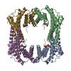
Mass: 18.015 Da / Num. of mol.: 5 / Source method: isolated from a natural source / Formula: H2O
Has ligand of interestN

-
Experimental details

-
Experiment

ExperimentMethod: X-RAY DIFFRACTION / Number of used crystals: 1

-
Sample preparation

CrystalDensity Matthews: 2.89 Å3/Da / Density % sol: 57.45 %
Crystal growTemperature: 277 K / Method: vapor diffusion, sitting drop
Details: 0.2M Sodium Fluoride, 0.1M bis-tris propane pH 6.5, 20% w/v PEG 3350

-
Data collection

DiffractionMean temperature: 100 K / Serial crystal experiment: N
Diffraction sourceSource: SYNCHROTRON / Site: ESRF / Beamline: ID23-1 / Wavelength: 0.972425 Å
DetectorType: DECTRIS PILATUS 6M / Detector: PIXEL / Date: Dec 1, 2022
RadiationProtocol: SINGLE WAVELENGTH / Monochromatic (M) / Laue (L): M / Scattering type: x-ray
Radiation wavelengthWavelength: 0.972425 Å / Relative weight: 1
ReflectionResolution: 2.5→137.59 Å / Num. obs: 43218 / % possible obs: 99.6 % / Redundancy: 3.8 % / Biso Wilson estimate: 59.6 Å2 / CC1/2: 0.996 / Net I/σ(I): 8.1
Reflection shellResolution: 2.5→2.59 Å / Redundancy: 3.6 % / Mean I/σ(I) obs: 1.3 / Num. unique obs: 4507 / CC1/2: 0.837

-
Processing

Software
NameVersionClassification
REFMAC5.8.0419refinement
autoPROCdata reduction
Aimlessdata scaling
MOLREPphasing
PDB_EXTRACTdata extraction
RefinementMethod to determine structure: MOLECULAR REPLACEMENT / Resolution: 2.5→137.59 Å / Cor.coef. Fo:Fc: 0.953 / Cor.coef. Fo:Fc free: 0.933 / SU B: 12.935 / SU ML: 0.271 / Cross valid method: THROUGHOUT / ESU R: 0.439 / ESU R Free: 0.281 / Stereochemistry target values: MAXIMUM LIKELIHOOD / Details: HYDROGENS HAVE BEEN ADDED IN THE RIDING POSITIONS
RfactorNum. reflection% reflectionSelection details
Rfree0.26001 2185 5.1 %RANDOM
Rwork0.21615 ---
obs0.21831 41031 99.56 %-
Solvent computationIon probe radii: 0.8 Å / Shrinkage radii: 0.8 Å / VDW probe radii: 1.2 Å / Solvent model: MASK
Displacement parametersBiso mean: 78.87 Å2
Baniso -1Baniso -2Baniso -3
1--2.02 Å20 Å24.95 Å2
2---3.68 Å20 Å2
3---1.07 Å2
Refinement stepCycle: 1 / Resolution: 2.5→137.59 Å
ProteinNucleic acidLigandSolventTotal
Num. atoms7225 0 0 5 7230
Refine LS restraints
Refine-IDTypeDev idealDev ideal targetNumber
X-RAY DIFFRACTIONr_bond_refined_d0.0050.0127485
X-RAY DIFFRACTIONr_bond_other_d0.0010.0166908
X-RAY DIFFRACTIONr_angle_refined_deg1.1271.65310173
X-RAY DIFFRACTIONr_angle_other_deg0.4011.56515922
X-RAY DIFFRACTIONr_dihedral_angle_1_deg5.5815991
X-RAY DIFFRACTIONr_dihedral_angle_2_deg7.529536
X-RAY DIFFRACTIONr_dihedral_angle_3_deg17.273101236
X-RAY DIFFRACTIONr_dihedral_angle_4_deg
X-RAY DIFFRACTIONr_chiral_restr0.0590.21175
X-RAY DIFFRACTIONr_gen_planes_refined0.0040.028879
X-RAY DIFFRACTIONr_gen_planes_other0.0010.021641
X-RAY DIFFRACTIONr_nbd_refined
X-RAY DIFFRACTIONr_nbd_other
X-RAY DIFFRACTIONr_nbtor_refined
X-RAY DIFFRACTIONr_nbtor_other
X-RAY DIFFRACTIONr_xyhbond_nbd_refined
X-RAY DIFFRACTIONr_xyhbond_nbd_other
X-RAY DIFFRACTIONr_metal_ion_refined
X-RAY DIFFRACTIONr_metal_ion_other
X-RAY DIFFRACTIONr_symmetry_vdw_refined
X-RAY DIFFRACTIONr_symmetry_vdw_other
X-RAY DIFFRACTIONr_symmetry_hbond_refined
X-RAY DIFFRACTIONr_symmetry_hbond_other
X-RAY DIFFRACTIONr_symmetry_metal_ion_refined
X-RAY DIFFRACTIONr_symmetry_metal_ion_other
X-RAY DIFFRACTIONr_mcbond_it6.1348.3433841
X-RAY DIFFRACTIONr_mcbond_other6.1338.3433841
X-RAY DIFFRACTIONr_mcangle_it8.86914.9674801
X-RAY DIFFRACTIONr_mcangle_other8.86814.9684802
X-RAY DIFFRACTIONr_scbond_it6.4488.6013644
X-RAY DIFFRACTIONr_scbond_other6.4478.6023645
X-RAY DIFFRACTIONr_scangle_it
X-RAY DIFFRACTIONr_scangle_other9.62915.6945349
X-RAY DIFFRACTIONr_long_range_B_refined11.56174.848416
X-RAY DIFFRACTIONr_long_range_B_other11.5674.858417
X-RAY DIFFRACTIONr_rigid_bond_restr
X-RAY DIFFRACTIONr_sphericity_free
X-RAY DIFFRACTIONr_sphericity_bonded
LS refinement shellResolution: 2.5→2.565 Å / Total num. of bins used: 20
RfactorNum. reflection% reflection
Rfree0.335 158 -
Rwork0.329 3028 -
obs--99.78 %

+
About Yorodumi

-
News

-
Feb 9, 2022. New format data for meta-information of EMDB entries

New format data for meta-information of EMDB entries

  • Version 3 of the EMDB header file is now the official format.
  • The previous official version 1.9 will be removed from the archive.

Related info.:EMDB header

External links:wwPDB to switch to version 3 of the EMDB data model

-
Aug 12, 2020. Covid-19 info

Covid-19 info

URL: https://pdbjlvh1.pdbj.org/emnavi/covid19.php

New page: Covid-19 featured information page in EM Navigator.

Related info.:Covid-19 info / Mar 5, 2020. Novel coronavirus structure data

+
Mar 5, 2020. Novel coronavirus structure data

Novel coronavirus structure data

Related info.:Yorodumi Speices / Aug 12, 2020. Covid-19 info

External links:COVID-19 featured content - PDBj / Molecule of the Month (242):Coronavirus Proteases

+
Jan 31, 2019. EMDB accession codes are about to change! (news from PDBe EMDB page)

EMDB accession codes are about to change! (news from PDBe EMDB page)

  • The allocation of 4 digits for EMDB accession codes will soon come to an end. Whilst these codes will remain in use, new EMDB accession codes will include an additional digit and will expand incrementally as the available range of codes is exhausted. The current 4-digit format prefixed with “EMD-” (i.e. EMD-XXXX) will advance to a 5-digit format (i.e. EMD-XXXXX), and so on. It is currently estimated that the 4-digit codes will be depleted around Spring 2019, at which point the 5-digit format will come into force.
  • The EM Navigator/Yorodumi systems omit the EMD- prefix.

Related info.:Q: What is EMD? / ID/Accession-code notation in Yorodumi/EM Navigator

External links:EMDB Accession Codes are Changing Soon! / Contact to PDBj

+
Jul 12, 2017. Major update of PDB

Major update of PDB

  • wwPDB released updated PDB data conforming to the new PDBx/mmCIF dictionary.
  • This is a major update changing the version number from 4 to 5, and with Remediation, in which all the entries are updated.
  • In this update, many items about electron microscopy experimental information are reorganized (e.g. em_software).
  • Now, EM Navigator and Yorodumi are based on the updated data.

External links:wwPDB Remediation / Enriched Model Files Conforming to OneDep Data Standards Now Available in the PDB FTP Archive

-
Yorodumi

Thousand views of thousand structures

  • Yorodumi is a browser for structure data from EMDB, PDB, SASBDB, etc.
  • This page is also the successor to EM Navigator detail page, and also detail information page/front-end page for Omokage search.
  • The word "yorodu" (or yorozu) is an old Japanese word meaning "ten thousand". "mi" (miru) is to see.

Related info.:EMDB / PDB / SASBDB / Comparison of 3 databanks / Yorodumi Search / Aug 31, 2016. New EM Navigator & Yorodumi / Yorodumi Papers / Jmol/JSmol / Function and homology information / Changes in new EM Navigator and Yorodumi

Read more