 Movie
Movie Controller


+ Open data

- Basic information
| Entry | Database: PDB / ID: 8qd3 | ||||||
|---|---|---|---|---|---|---|---|
| Title | Ayg1p in complex with 1,3,6,8-Tetrahydroxynaphthalene | ||||||
 Components Components | Pigment biosynthesis protein yellowish-green 1 | ||||||
 Keywords Keywords | LYASE / Natural Product Biosynthesis / Polyketide Synthase System / alpha / beta-Hydrolase Fold / Polyketide Shortening / Retro Claisen Reaction | ||||||
| Function / homology | Esterase FrsA-like / Esterase FrsA-like / : / Alpha/Beta hydrolase fold / hydrolase activity / FLAVIOLIN / TRIETHYLENE GLYCOL / : / Heptaketide hydrolyase ayg1 Function and homology information Function and homology information | ||||||
| Biological species |  | ||||||
| Method |  X-RAY DIFFRACTION / SYNCHROTRON / MOLECULAR REPLACEMENT / Resolution: 1.7 Å X-RAY DIFFRACTION / SYNCHROTRON / MOLECULAR REPLACEMENT / Resolution: 1.7 Å | ||||||
 Authors Authors | Schmalhofer, M. / Vagstad, A.L. / Zhou, Q. / Bode, H.B. / Groll, M. | ||||||
| Funding support |  Germany, 1items Germany, 1items
| ||||||
 Citation Citation | Journal: Adv Sci / Year: 2024 Title: Polyketide Trimming Shapes Dihydroxynaphthalene-Melanin and Anthraquinone Pigments. Authors: Schmalhofer, M. / Vagstad, A.L. / Zhou, Q. / Bode, H.B. / Groll, M. | ||||||
| History |
|
- Structure visualization
| Structure viewer | Molecule: MolmilJmol/JSmol |
|---|
- Downloads & links
-Download
| PDBx/mmCIF format | 8qd3.cif.gz | 663.4 KB | Display | PDBx/mmCIF format |
|---|---|---|---|---|
| PDB format | pdb8qd3.ent.gz | 551.9 KB | Display | PDB format |
| PDBx/mmJSON format | 8qd3.json.gz | Tree view | PDBx/mmJSON format | |
| Others |  Other downloads Other downloads |
-Validation report
| Arichive directory | https://data.pdbj.org/pub/pdb/validation_reports/qd/8qd3ftp://data.pdbj.org/pub/pdb/validation_reports/qd/8qd3 | HTTPS FTP |
|---|
-Related structure data
| Related structure data | 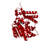 8qbhC  8qbiC  8qd1C  8qd2C  8qd4C  8qd5C  8qd6C  8qd7C  8qd8C  8qd9C  8qdaC  8qdbC C: citing same article ( |
|---|---|
| Similar structure data |

-Links
 PDBj
PDBj- Assembly
| Deposited unit | 
| ||||||||
|---|---|---|---|---|---|---|---|---|---|
| 1 | 
| ||||||||
| 2 | 
| ||||||||
| 3 | 
| ||||||||
| 4 | 
| ||||||||
| Unit cell |
|
-Components
-Protein , 1 types, 4 molecules ABCD
| #1: Protein | Mass: 47440.410 Da / Num. of mol.: 4 Source method: isolated from a genetically manipulated source Source: (gene. exp.)  |
|---|
-Non-polymers , 5 types, 1043 molecules 
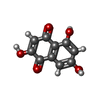


| #2: Chemical | ChemComp-QW8 / Mass: 192.168 Da / Num. of mol.: 4 / Source method: obtained synthetically / Formula: C10H8O4 / Feature type: SUBJECT OF INVESTIGATION #3: Chemical |  Mass: 150.173 Da / Num. of mol.: 2 / Source method: obtained synthetically / Formula: C6H14O4 Mass: 150.173 Da / Num. of mol.: 2 / Source method: obtained synthetically / Formula: C6H14O4#4: Chemical | ChemComp-FLV /  Mass: 206.152 Da / Num. of mol.: 8 / Source method: obtained synthetically / Formula: C10H6O5 Mass: 206.152 Da / Num. of mol.: 8 / Source method: obtained synthetically / Formula: C10H6O5#5: Chemical |  Mass: 62.068 Da / Num. of mol.: 2 / Source method: obtained synthetically / Formula: C2H6O2 Mass: 62.068 Da / Num. of mol.: 2 / Source method: obtained synthetically / Formula: C2H6O2#6: Water | ChemComp-HOH / | Mass: 18.015 Da / Num. of mol.: 1027 / Source method: isolated from a natural source / Formula: H2O |
|---|
-Details
| Has ligand of interest | Y |
|---|
-Experimental details
-Experiment
| Experiment | Method: X-RAY DIFFRACTION / Number of used crystals: 1 |
|---|
- Sample preparation
| Crystal | Density Matthews: 2.24 Å3/Da / Density % sol: 45.18 % |
|---|---|
| Crystal grow | Temperature: 293 K / Method: vapor diffusion, hanging drop / Details: 100 mM K/Na tartrate, 25.5% PEG 3350 |
-Data collection
| Diffraction | Mean temperature: 100 K / Serial crystal experiment: N |
|---|---|
| Diffraction source | Source: SYNCHROTRON / Site: SLS  / Beamline: X06SA / Wavelength: 1 Å / Beamline: X06SA / Wavelength: 1 Å |
| Detector | Type: DECTRIS EIGER X 16M / Detector: PIXEL / Date: Nov 27, 2021 |
| Radiation | Protocol: SINGLE WAVELENGTH / Monochromatic (M) / Laue (L): M / Scattering type: x-ray |
| Radiation wavelength | Wavelength: 1 Å / Relative weight: 1 |
| Reflection | Resolution: 1.7→50 Å / Num. obs: 173708 / % possible obs: 94.6 % / Redundancy: 3.2 % / Rmerge(I) obs: 0.094 / Net I/σ(I): 6.7 |
| Reflection shell | Resolution: 1.7→1.8 Å / Rmerge(I) obs: 0.6 / Mean I/σ(I) obs: 1.9 / Num. unique obs: 27645 / % possible all: 95.7 |
- Processing
| Software |
| ||||||||||||||||||||||||||||||||||||||||||||||||||||||||||||||||||||||||||||||||||||||||||||||||||||||||||||||||||||||||||||||||||||||||||||||||||||||||||||||||||||||||||||||||||||||
|---|---|---|---|---|---|---|---|---|---|---|---|---|---|---|---|---|---|---|---|---|---|---|---|---|---|---|---|---|---|---|---|---|---|---|---|---|---|---|---|---|---|---|---|---|---|---|---|---|---|---|---|---|---|---|---|---|---|---|---|---|---|---|---|---|---|---|---|---|---|---|---|---|---|---|---|---|---|---|---|---|---|---|---|---|---|---|---|---|---|---|---|---|---|---|---|---|---|---|---|---|---|---|---|---|---|---|---|---|---|---|---|---|---|---|---|---|---|---|---|---|---|---|---|---|---|---|---|---|---|---|---|---|---|---|---|---|---|---|---|---|---|---|---|---|---|---|---|---|---|---|---|---|---|---|---|---|---|---|---|---|---|---|---|---|---|---|---|---|---|---|---|---|---|---|---|---|---|---|---|---|---|---|---|
| Refinement | Method to determine structure: MOLECULAR REPLACEMENT / Resolution: 1.7→46.48 Å / Cor.coef. Fo:Fc: 0.957 / Cor.coef. Fo:Fc free: 0.95 / SU B: 3.265 / SU ML: 0.053 / Cross valid method: THROUGHOUT / ESU R: 0.042 / ESU R Free: 0.024 / Stereochemistry target values: MAXIMUM LIKELIHOOD / Details: HYDROGENS HAVE BEEN ADDED IN THE RIDING POSITIONS
| ||||||||||||||||||||||||||||||||||||||||||||||||||||||||||||||||||||||||||||||||||||||||||||||||||||||||||||||||||||||||||||||||||||||||||||||||||||||||||||||||||||||||||||||||||||||
| Solvent computation | Ion probe radii: 0.8 Å / Shrinkage radii: 0.8 Å / VDW probe radii: 1.2 Å / Solvent model: MASK | ||||||||||||||||||||||||||||||||||||||||||||||||||||||||||||||||||||||||||||||||||||||||||||||||||||||||||||||||||||||||||||||||||||||||||||||||||||||||||||||||||||||||||||||||||||||
| Displacement parameters | Biso mean: 20.791 Å2
| ||||||||||||||||||||||||||||||||||||||||||||||||||||||||||||||||||||||||||||||||||||||||||||||||||||||||||||||||||||||||||||||||||||||||||||||||||||||||||||||||||||||||||||||||||||||
| Refinement step | Cycle: 1 / Resolution: 1.7→46.48 Å
| ||||||||||||||||||||||||||||||||||||||||||||||||||||||||||||||||||||||||||||||||||||||||||||||||||||||||||||||||||||||||||||||||||||||||||||||||||||||||||||||||||||||||||||||||||||||
| Refine LS restraints |
|