 Movie
Movie Controller

[English] 日本語
 Yorodumi
- PDB-8qck: Crystal structure of mycothiol disulfide reductase Mtr from Mycob... -
+ Open data

- Basic information
| Entry | Database: PDB / ID: 8qck | ||||||
|---|---|---|---|---|---|---|---|
| Title | Crystal structure of mycothiol disulfide reductase Mtr from Mycobacterium smegmatis | ||||||
 Components Components | Pyridine nucleotide-disulfide oxidoreductase dimerization region | ||||||
 Keywords Keywords | OXIDOREDUCTASE / DISULFIDE REDUCTASE / FLAVOPROTEIN / MYCOTHIONE REDUCTASE | ||||||
| Function / homology |  Function and homology information Function and homology informationOxidoreductases; Acting on a sulfur group of donors; With NAD+ or NADP+ as acceptor / oxidoreductase activity, acting on a sulfur group of donors, NAD(P) as acceptor / nucleotide binding Similarity search - Function | ||||||
| Biological species |  Mycolicibacterium smegmatis MC2 155 (bacteria) Mycolicibacterium smegmatis MC2 155 (bacteria) | ||||||
| Method |  X-RAY DIFFRACTION / SYNCHROTRON / MOLECULAR REPLACEMENT / Resolution: 4.7 Å X-RAY DIFFRACTION / SYNCHROTRON / MOLECULAR REPLACEMENT / Resolution: 4.7 Å | ||||||
 Authors Authors | Gutierrez-Fernandez, J. / Hammerstad, M. / Hersleth, H.-P. | ||||||
| Funding support |  Norway, 1items Norway, 1items
| ||||||
 Citation Citation | Journal: Acta Crystallogr D Struct Biol / Year: 2024 Title: The crystal structure of mycothiol disulfide reductase (Mtr) provides mechanistic insight into the specific low-molecular-weight thiol reductase activity of Actinobacteria. Authors: Gutierrez-Fernandez, J. / Hersleth, H.P. / Hammerstad, M. | ||||||
| History |
|
- Structure visualization
| Structure viewer | Molecule: MolmilJmol/JSmol |
|---|
- Downloads & links
-Download
| PDBx/mmCIF format | 8qck.cif.gz | 225.8 KB | Display | PDBx/mmCIF format |
|---|---|---|---|---|
| PDB format | pdb8qck.ent.gz | 146.3 KB | Display | PDB format |
| PDBx/mmJSON format | 8qck.json.gz | Tree view | PDBx/mmJSON format | |
| Others |  Other downloads Other downloads |
-Validation report
| Arichive directory | https://data.pdbj.org/pub/pdb/validation_reports/qc/8qckftp://data.pdbj.org/pub/pdb/validation_reports/qc/8qck | HTTPS FTP |
|---|
-Related structure data
| Related structure data |  8qcjSC S: Starting model for refinement C: citing same article ( |
|---|---|
| Similar structure data |
-Links
 PDBj
PDBj


- Assembly
| Deposited unit | 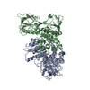
| ||||||||||||||||||
|---|---|---|---|---|---|---|---|---|---|---|---|---|---|---|---|---|---|---|---|
| 1 |
| ||||||||||||||||||
| Unit cell |
| ||||||||||||||||||
| Noncrystallographic symmetry (NCS) | NCS domain:
NCS domain segments: Component-ID: 1 / Ens-ID: ens_1 / Beg auth comp-ID: MET / Beg label comp-ID: MET / End auth comp-ID: GLU / End label comp-ID: GLU / Auth seq-ID: 1 - 461 / Label seq-ID: 1 - 461
NCS oper: (Code: givenMatrix: (-0.979197738181, -0.00656999600635, 0.202801934641), (0.00495677867763, -0.999951915315, -0.00846152490312), (0.202847775158, -0.00728026174129, 0.979183219782)Vector: ...NCS oper: (Code: given Matrix: (-0.979197738181, -0.00656999600635, 0.202801934641), Vector: |
-Components
| #1: Protein | Mass: 49998.836 Da / Num. of mol.: 2 Source method: isolated from a genetically manipulated source Source: (gene. exp.) Mycolicibacterium smegmatis MC2 155 (bacteria)Gene: gorA, MSMEI_2549 / Production host: References: UniProt: I7G0D4, Oxidoreductases; Acting on a sulfur group of donors; With NAD+ or NADP+ as acceptor |
|---|
-Experimental details
-Experiment
| Experiment | Method: X-RAY DIFFRACTION / Number of used crystals: 1 |
|---|
- Sample preparation
| Crystal | Density Matthews: 3.3 Å3/Da / Density % sol: 62.9 % |
|---|---|
| Crystal grow | Temperature: 293 K / Method: vapor diffusion, sitting drop / pH: 7.5 Details: 0.02 M magnesium chloride hexahydrate, 0.1 M HEPES pH 7.5 and 22% (w/v) poly-(acrylic acid sodium salt) 5,100, and a protein concentration of 12.5 mg/mL. |
-Data collection
| Diffraction | Mean temperature: 100 K / Serial crystal experiment: N |
|---|---|
| Diffraction source | Source: SYNCHROTRON / Site: ESRF  / Beamline: ID30B / Wavelength: 0.9184 Å / Beamline: ID30B / Wavelength: 0.9184 Å |
| Detector | Type: DECTRIS EIGER2 X 9M / Detector: PIXEL / Date: Oct 5, 2022 |
| Radiation | Protocol: SINGLE WAVELENGTH / Monochromatic (M) / Laue (L): M / Scattering type: x-ray |
| Radiation wavelength | Wavelength: 0.9184 Å / Relative weight: 1 |
| Reflection | Resolution: 4.7→49.4 Å / Num. obs: 7284 / % possible obs: 99.9 % / Redundancy: 6.1 % / Biso Wilson estimate: 55.63 Å2 / CC1/2: 0.942 / Rmerge(I) obs: 0.635 / Rpim(I) all: 0.278 / Rrim(I) all: 0.695 / Net I/σ(I): 3 |
| Reflection shell | Resolution: 4.7→5.25 Å / Redundancy: 6.3 % / Rmerge(I) obs: 1.183 / Mean I/σ(I) obs: 1.6 / Num. unique obs: 2015 / CC1/2: 0.847 / Rpim(I) all: 0.514 / Rrim(I) all: 1.292 / % possible all: 99.9 |
- Processing
| Software |
| ||||||||||||||||||||||||||||||||||||||||||
|---|---|---|---|---|---|---|---|---|---|---|---|---|---|---|---|---|---|---|---|---|---|---|---|---|---|---|---|---|---|---|---|---|---|---|---|---|---|---|---|---|---|---|---|
| Refinement | Method to determine structure: MOLECULAR REPLACEMENT Starting model: 8QCJ Resolution: 4.7→49.37 Å / SU ML: 0.8538 / Cross valid method: FREE R-VALUE / σ(F): 1.33 / Phase error: 41.1901 Stereochemistry target values: GeoStd + Monomer Library + CDL v1.2
| ||||||||||||||||||||||||||||||||||||||||||
| Solvent computation | Shrinkage radii: 0.9 Å / VDW probe radii: 1.1 Å / Solvent model: FLAT BULK SOLVENT MODEL | ||||||||||||||||||||||||||||||||||||||||||
| Displacement parameters | Biso mean: 113.65 Å2 | ||||||||||||||||||||||||||||||||||||||||||
| Refine analyze | Luzzati coordinate error obs: 1.243 Å | ||||||||||||||||||||||||||||||||||||||||||
| Refinement step | Cycle: LAST / Resolution: 4.7→49.37 Å
| ||||||||||||||||||||||||||||||||||||||||||
| Refine LS restraints |
| ||||||||||||||||||||||||||||||||||||||||||
| Refine LS restraints NCS | Type: Torsion NCS / Rms dev position: 1.39684432767 Å | ||||||||||||||||||||||||||||||||||||||||||
| LS refinement shell |
|
