| Entry | Database: PDB / ID: 8q6h
|
|---|
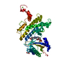
| Title | HUMAN PI4KIIIB IN COMPLEX WITH COVALENTLY BOUND INHIBITOR (COMPOUND 11) |
|---|
 Components Components | Phosphatidylinositol 4-kinase beta |
|---|
 Keywords Keywords | TRANSFERASE / LIPID KINASE / TRANSFERASE-SIGNALING PROTEIN COMPLEX / COVALENT INHIBITOR. |
|---|
| Function / homology |  Function and homology information Function and homology information
1-phosphatidylinositol 4-kinase / 1-phosphatidylinositol 4-kinase activity / rough endoplasmic reticulum membrane / Synthesis of PIPs at the Golgi membrane / phosphatidylinositol biosynthetic process / lysosome organization / phosphatidylinositol-mediated signaling / phosphatidylinositol phosphate biosynthetic process / inner ear development / 14-3-3 protein binding ...1-phosphatidylinositol 4-kinase / 1-phosphatidylinositol 4-kinase activity / rough endoplasmic reticulum membrane / Synthesis of PIPs at the Golgi membrane / phosphatidylinositol biosynthetic process / lysosome organization / phosphatidylinositol-mediated signaling / phosphatidylinositol phosphate biosynthetic process / inner ear development / 14-3-3 protein binding / receptor-mediated endocytosis / mitochondrial outer membrane / endosome / Golgi membrane / perinuclear region of cytoplasm / Golgi apparatus / signal transduction / ATP binding / membrane / cytoplasm / cytosolSimilarity search - Function : / PI4KB/PIK1, accessory (PIK) domain / Phosphoinositide 3-kinase, accessory (PIK) domain / Phosphatidylinositol kinase / PIK helical domain profile. / Phosphatidylinositol 3- and 4-kinases signature 1. / Phosphatidylinositol 3/4-kinase, conserved site / Phosphatidylinositol 3- and 4-kinases signature 2. / Phosphatidylinositol 3-/4-kinase, catalytic domain superfamily / Phosphoinositide 3-kinase, catalytic domain ...: / PI4KB/PIK1, accessory (PIK) domain / Phosphoinositide 3-kinase, accessory (PIK) domain / Phosphatidylinositol kinase / PIK helical domain profile. / Phosphatidylinositol 3- and 4-kinases signature 1. / Phosphatidylinositol 3/4-kinase, conserved site / Phosphatidylinositol 3- and 4-kinases signature 2. / Phosphatidylinositol 3-/4-kinase, catalytic domain superfamily / Phosphoinositide 3-kinase, catalytic domain / Phosphatidylinositol 3- and 4-kinase / Phosphatidylinositol 3- and 4-kinases catalytic domain profile. / Phosphatidylinositol 3-/4-kinase, catalytic domain / Protein kinase-like domain superfamilySimilarity search - Domain/homology |
|---|
| Biological species |  Homo sapiens (human) Homo sapiens (human) |
|---|
| Method |  X-RAY DIFFRACTION / SYNCHROTRON / FOURIER SYNTHESIS / Resolution: 1.94 Å X-RAY DIFFRACTION / SYNCHROTRON / FOURIER SYNTHESIS / Resolution: 1.94 Å |
|---|
 Authors Authors | Somers, D.O. |
|---|
| Funding support | 1items | Organization | Grant number | Country |
|---|
| Not funded | | |
|
|---|
 Citation Citation | Journal: Rsc Chem Biol / Year: 2023
Title: Covalent targeting of non-cysteine residues in PI4KIII beta.
Authors: Cosgrove, B. / Grant, E.K. / Bertrand, S. / Down, K.D. / Somers, D.O. / P Evans, J. / Tomkinson, N.C.O. / Barker, M.D. |
|---|
| History | | Deposition | Aug 11, 2023 | Deposition site: PDBE / Processing site: PDBE |
|---|
| Revision 1.0 | Dec 13, 2023 | Provider: repository / Type: Initial release |
|---|
| Revision 1.1 | Nov 13, 2024 | Group: Structure summary / Category: pdbx_entry_details / pdbx_modification_feature / Item: _pdbx_entry_details.has_protein_modification |
|---|
|
|---|
 Movie
Movie Controller

 Yorodumi
 Open data
 Basic information
 Structure visualization
 Downloads & links
 Other downloads
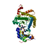
 PDBj
PDBj
 Assembly
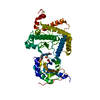
 Baculovirus expression vector pFastBac1-HM
 Mass: 24.305 Da / Num. of mol.: 1 / Source method: obtained synthetically / Formula: Mg
 Mass: 554.544 Da / Num. of mol.: 1 / Source method: obtained synthetically / Formula: C22H20F2N4O7S2 / Feature type: SUBJECT OF INVESTIGATION
 Mass: 62.068 Da / Num. of mol.: 1 / Source method: obtained synthetically / Formula: C2H6O2
 Mass: 18.015 Da / Num. of mol.: 249 / Source method: isolated from a natural source / Formula: H2O
 Sample preparation
 / Beamline: I04 / Wavelength: 0.9795 Å
 Processing