 Movie
Movie Controller

[English] 日本語
 Yorodumi
- PDB-8q5a: Crystal structure of metal-dependent class II sulfofructosephosph... -
+ Open data

- Basic information
| Entry | Database: PDB / ID: 8q5a | ||||||
|---|---|---|---|---|---|---|---|
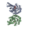
| Title | Crystal structure of metal-dependent class II sulfofructosephosphate aldolase from Hafnia paralvei HpSqiA-Zn in complex with dihydroxyacetone phosphate (DHAP) | ||||||
 Components Components | Ketose-bisphosphate aldolase | ||||||
 Keywords Keywords | LYASE / metal-dependent / aldolase / sulfoquinovose / sulfofructose phosphate | ||||||
| Function / homology |  Function and homology information Function and homology informationaldehyde-lyase activity / carbohydrate metabolic process / zinc ion binding Similarity search - Function | ||||||
| Biological species |  Hafnia paralvei (bacteria) Hafnia paralvei (bacteria) | ||||||
| Method |  X-RAY DIFFRACTION / SYNCHROTRON / MOLECULAR REPLACEMENT / Resolution: 2.8 Å X-RAY DIFFRACTION / SYNCHROTRON / MOLECULAR REPLACEMENT / Resolution: 2.8 Å | ||||||
 Authors Authors | Sharma, M. / Davies, G.J. | ||||||
| Funding support |  United Kingdom, 1items United Kingdom, 1items
| ||||||
 Citation Citation | Journal: J.Biol.Chem. / Year: 2023 Title: Defining the molecular architecture, metal dependence, and distribution of metal-dependent class II sulfofructose-1-phosphate aldolases. Authors: Sharma, M. / Kaur, A. / Madiedo Soler, N. / Lingford, J.P. / Epa, R. / Goddard-Borger, E.D. / Davies, G.J. / Williams, S.J. | ||||||
| History |
|
- Structure visualization
| Structure viewer | Molecule: MolmilJmol/JSmol |
|---|
- Downloads & links
-Download
| PDBx/mmCIF format | 8q5a.cif.gz | 68.5 KB | Display | PDBx/mmCIF format |
|---|---|---|---|---|
| PDB format | pdb8q5a.ent.gz | 46.1 KB | Display | PDB format |
| PDBx/mmJSON format | 8q5a.json.gz | Tree view | PDBx/mmJSON format | |
| Others |  Other downloads Other downloads |
-Validation report
| Arichive directory | https://data.pdbj.org/pub/pdb/validation_reports/q5/8q5aftp://data.pdbj.org/pub/pdb/validation_reports/q5/8q5a | HTTPS FTP |
|---|
-Related structure data


-Links
 PDBj
PDBj
- Assembly
| Deposited unit | 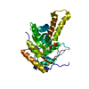
| ||||||||
|---|---|---|---|---|---|---|---|---|---|
| 1 | 
| ||||||||
| Unit cell |
| ||||||||
| Components on special symmetry positions |
|
-Components
| #1: Protein | Mass: 33588.234 Da / Num. of mol.: 1 Source method: isolated from a genetically manipulated source Source: (gene. exp.) Hafnia paralvei (bacteria) / Gene: kbaY / Plasmid: pET28a (+) / Production host: | ||||||
|---|---|---|---|---|---|---|---|
| #2: Chemical |   Mass: 65.409 Da / Num. of mol.: 2 / Source method: obtained synthetically / Formula: Zn / Feature type: SUBJECT OF INVESTIGATION Mass: 65.409 Da / Num. of mol.: 2 / Source method: obtained synthetically / Formula: Zn / Feature type: SUBJECT OF INVESTIGATION#3: Chemical | ChemComp-13P / |   Mass: 170.058 Da / Num. of mol.: 1 / Source method: obtained synthetically / Formula: C3H7O6P / Feature type: SUBJECT OF INVESTIGATION Mass: 170.058 Da / Num. of mol.: 1 / Source method: obtained synthetically / Formula: C3H7O6P / Feature type: SUBJECT OF INVESTIGATION#4: Water | ChemComp-HOH / |  Mass: 18.015 Da / Num. of mol.: 6 / Source method: isolated from a natural source / Formula: H2O Mass: 18.015 Da / Num. of mol.: 6 / Source method: isolated from a natural source / Formula: H2OHas ligand of interest | Y | |
-Experimental details
-Experiment
| Experiment | Method: X-RAY DIFFRACTION / Number of used crystals: 1 |
|---|
- Sample preparation
| Crystal | Density Matthews: 2.44 Å3/Da / Density % sol: 49.58 % |
|---|---|
| Crystal grow | Temperature: 293 K / Method: vapor diffusion, sitting drop Details: A crystal of the DHAP complex grew from 14 mg mL-1 enzyme in 50 mM Tris buffer pH 7.4 in a drop with 0.15 uL protein to 0.15 uL mother liquor soaked with SFP for 2 min, with the reservoir ...Details: A crystal of the DHAP complex grew from 14 mg mL-1 enzyme in 50 mM Tris buffer pH 7.4 in a drop with 0.15 uL protein to 0.15 uL mother liquor soaked with SFP for 2 min, with the reservoir solution containing 25% PEG 1,500 w/v, 0.1 M SPG (succinic acid, propionate, glycine) buffer pH 7 |
-Data collection
| Diffraction | Mean temperature: 100 K / Serial crystal experiment: N |
|---|---|
| Diffraction source | Source: SYNCHROTRON / Site: Diamond / Beamline: I03 / Wavelength: 0.97625 Å |
| Detector | Type: DECTRIS PILATUS 2M / Detector: PIXEL / Date: Aug 10, 2020 |
| Radiation | Protocol: SINGLE WAVELENGTH / Monochromatic (M) / Laue (L): M / Scattering type: x-ray |
| Radiation wavelength | Wavelength: 0.97625 Å / Relative weight: 1 |
| Reflection | Resolution: 2.8→34.22 Å / Num. obs: 7522 / % possible obs: 99.9 % / Redundancy: 12.7 % / CC1/2: 0.997 / Rmerge(I) obs: 0.101 / Rpim(I) all: 0.029 / Rrim(I) all: 0.105 / Χ2: 0.99 / Net I/σ(I): 17.7 / Num. measured all: 95222 |
| Reflection shell | Resolution: 2.8→2.95 Å / % possible obs: 100 % / Redundancy: 13.4 % / Rmerge(I) obs: 0.439 / Num. measured all: 14614 / Num. unique obs: 1087 / CC1/2: 0.971 / Rpim(I) all: 0.123 / Rrim(I) all: 0.456 / Χ2: 0.95 / Net I/σ(I) obs: 5.6 |
- Processing
| Software |
| ||||||||||||||||||||||||||||||||||||||||||||||||||||||||||||||||||||||||||||||||||||||||||||||||||||||||||||||||||||||||||||||||||||||||||||||||||||||||||||||||||||||||||||||||||||||
|---|---|---|---|---|---|---|---|---|---|---|---|---|---|---|---|---|---|---|---|---|---|---|---|---|---|---|---|---|---|---|---|---|---|---|---|---|---|---|---|---|---|---|---|---|---|---|---|---|---|---|---|---|---|---|---|---|---|---|---|---|---|---|---|---|---|---|---|---|---|---|---|---|---|---|---|---|---|---|---|---|---|---|---|---|---|---|---|---|---|---|---|---|---|---|---|---|---|---|---|---|---|---|---|---|---|---|---|---|---|---|---|---|---|---|---|---|---|---|---|---|---|---|---|---|---|---|---|---|---|---|---|---|---|---|---|---|---|---|---|---|---|---|---|---|---|---|---|---|---|---|---|---|---|---|---|---|---|---|---|---|---|---|---|---|---|---|---|---|---|---|---|---|---|---|---|---|---|---|---|---|---|---|---|
| Refinement | Method to determine structure: MOLECULAR REPLACEMENT / Resolution: 2.8→34 Å / Cor.coef. Fo:Fc: 0.93 / Cor.coef. Fo:Fc free: 0.895 / SU B: 13.866 / SU ML: 0.281 / Cross valid method: THROUGHOUT / ESU R Free: 0.412 / Stereochemistry target values: MAXIMUM LIKELIHOOD / Details: HYDROGENS HAVE BEEN ADDED IN THE RIDING POSITIONS
| ||||||||||||||||||||||||||||||||||||||||||||||||||||||||||||||||||||||||||||||||||||||||||||||||||||||||||||||||||||||||||||||||||||||||||||||||||||||||||||||||||||||||||||||||||||||
| Solvent computation | Ion probe radii: 0.8 Å / Shrinkage radii: 0.8 Å / VDW probe radii: 1.2 Å / Solvent model: MASK | ||||||||||||||||||||||||||||||||||||||||||||||||||||||||||||||||||||||||||||||||||||||||||||||||||||||||||||||||||||||||||||||||||||||||||||||||||||||||||||||||||||||||||||||||||||||
| Displacement parameters | Biso mean: 46.956 Å2
| ||||||||||||||||||||||||||||||||||||||||||||||||||||||||||||||||||||||||||||||||||||||||||||||||||||||||||||||||||||||||||||||||||||||||||||||||||||||||||||||||||||||||||||||||||||||
| Refinement step | Cycle: 1 / Resolution: 2.8→34 Å
| ||||||||||||||||||||||||||||||||||||||||||||||||||||||||||||||||||||||||||||||||||||||||||||||||||||||||||||||||||||||||||||||||||||||||||||||||||||||||||||||||||||||||||||||||||||||
| Refine LS restraints |
|