 Movie
Movie Controller

[English] 日本語
 Yorodumi
- PDB-8q1k: Structural analysis of PLD3 reveals insights into the mechanism o... -
+ Open data

- Basic information
| Entry | Database: PDB / ID: 8q1k | ||||||
|---|---|---|---|---|---|---|---|
| Title | Structural analysis of PLD3 reveals insights into the mechanism of lysosomal 5' exonuclease-mediated nucleic acid degradation | ||||||
 Components Components | 5'-3' exonuclease PLD3 | ||||||
 Keywords Keywords | DNA BINDING PROTEIN / PLD3 / structural biology / lysosome / DNA/RNA degradation / phospholipase D / 5' exonuclease | ||||||
| Function / homology |  Function and homology information Function and homology informationspleen exonuclease / Synthesis of PG / single-stranded DNA 5'-3' DNA exonuclease activity / myotube differentiation / Hydrolases; Acting on ester bonds; Phosphoric-diester hydrolases / D-type glycerophospholipase activity / regulation of cytokine production involved in inflammatory response / immune system process / Role of phospholipids in phagocytosis / lysosomal lumen ...spleen exonuclease / Synthesis of PG / single-stranded DNA 5'-3' DNA exonuclease activity / myotube differentiation / Hydrolases; Acting on ester bonds; Phosphoric-diester hydrolases / D-type glycerophospholipase activity / regulation of cytokine production involved in inflammatory response / immune system process / Role of phospholipids in phagocytosis / lysosomal lumen / lipid metabolic process / late endosome membrane / early endosome membrane / inflammatory response / Golgi membrane / lysosomal membrane / endoplasmic reticulum membrane / extracellular exosome Similarity search - Function | ||||||
| Biological species |  Homo sapiens (human) Homo sapiens (human) | ||||||
| Method |  X-RAY DIFFRACTION / SYNCHROTRON / SAD / Resolution: 1.51 Å X-RAY DIFFRACTION / SYNCHROTRON / SAD / Resolution: 1.51 Å | ||||||
 Authors Authors | Roske, Y. / Daumke, O. / Damme, M. | ||||||
| Funding support |  Germany, 1items Germany, 1items
| ||||||
 Citation Citation | Journal: Nucleic Acids Res. / Year: 2024 Title: Structural analysis of PLD3 reveals insights into the mechanism of lysosomal 5' exonuclease-mediated nucleic acid degradation. Authors: Roske, Y. / Cappel, C. / Cremer, N. / Hoffmann, P. / Koudelka, T. / Tholey, A. / Heinemann, U. / Daumke, O. / Damme, M. | ||||||
| History |
|
- Structure visualization
| Structure viewer | Molecule: MolmilJmol/JSmol |
|---|
- Downloads & links
-Download
| PDBx/mmCIF format | 8q1k.cif.gz | 418.7 KB | Display | PDBx/mmCIF format |
|---|---|---|---|---|
| PDB format | pdb8q1k.ent.gz | 339.5 KB | Display | PDB format |
| PDBx/mmJSON format | 8q1k.json.gz | Tree view | PDBx/mmJSON format | |
| Others |  Other downloads Other downloads |
-Validation report
| Arichive directory | https://data.pdbj.org/pub/pdb/validation_reports/q1/8q1kftp://data.pdbj.org/pub/pdb/validation_reports/q1/8q1k | HTTPS FTP |
|---|
-Related structure data

-Links
 PDBj
PDBj


- Assembly
| Deposited unit | 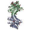
| ||||||||
|---|---|---|---|---|---|---|---|---|---|
| 1 |
| ||||||||
| Unit cell |
|
-Components
-Protein , 1 types, 2 molecules AB
| #1: Protein | Mass: 46945.766 Da / Num. of mol.: 2 Source method: isolated from a genetically manipulated source Source: (gene. exp.) Homo sapiens (human) / Gene: PLD3 / Production host:  |
|---|
-Sugars , 3 types, 4 molecules
| #2: Polysaccharide | alpha-D-mannopyranose-(1-3)-[alpha-D-mannopyranose-(1-6)]alpha-D-mannopyranose-(1-6)-[alpha-D- ...alpha-D-mannopyranose-(1-3)-[alpha-D-mannopyranose-(1-6)]alpha-D-mannopyranose-(1-6)-[alpha-D-mannopyranose-(1-3)]alpha-D-mannopyranose-(1-4)-2-acetamido-2-deoxy-beta-D-glucopyranose-(1-4)-2-acetamido-2-deoxy-beta-D-glucopyranose Type: oligosaccharide / Mass: 1235.105 Da / Num. of mol.: 1 Source method: isolated from a genetically manipulated source | ||
|---|---|---|---|
| #3: Polysaccharide | Source method: isolated from a genetically manipulated source #4: Polysaccharide | alpha-D-mannopyranose-(1-3)-[alpha-D-mannopyranose-(1-6)]alpha-D-mannopyranose-(1-6)-[alpha-D- ...alpha-D-mannopyranose-(1-3)-[alpha-D-mannopyranose-(1-6)]alpha-D-mannopyranose-(1-6)-[alpha-D-mannopyranose-(1-3)]beta-D-mannopyranose-(1-4)-2-acetamido-2-deoxy-beta-D-glucopyranose-(1-4)-2-acetamido-2-deoxy-beta-D-glucopyranose | |
-Non-polymers , 4 types, 1243 molecules 


| #5: Chemical | ChemComp-EDO /  Mass: 62.068 Da / Num. of mol.: 7 / Source method: obtained synthetically / Formula: C2H6O2 Mass: 62.068 Da / Num. of mol.: 7 / Source method: obtained synthetically / Formula: C2H6O2#6: Chemical | ChemComp-PO4 /  Mass: 94.971 Da / Num. of mol.: 8 / Source method: obtained synthetically / Formula: PO4 Mass: 94.971 Da / Num. of mol.: 8 / Source method: obtained synthetically / Formula: PO4#7: Chemical | ChemComp-MN /  Mass: 54.938 Da / Num. of mol.: 4 / Source method: isolated from a natural source / Formula: Mn Mass: 54.938 Da / Num. of mol.: 4 / Source method: isolated from a natural source / Formula: Mn#8: Water | ChemComp-HOH / | Mass: 18.015 Da / Num. of mol.: 1224 / Source method: isolated from a natural source / Formula: H2O |
|---|
-Details
| Has ligand of interest | N |
|---|
-Experimental details
-Experiment
| Experiment | Method: X-RAY DIFFRACTION / Number of used crystals: 1 |
|---|
- Sample preparation
| Crystal grow | Temperature: 293 K / Method: vapor diffusion, sitting drop / Details: 17% PEG 4000, 0.02 M NaI, 0.6 M Ammonium sulfate |
|---|
-Data collection
| Diffraction | Mean temperature: 100 K / Serial crystal experiment: N |
|---|---|
| Diffraction source | Source: SYNCHROTRON / Site: BESSY / Beamline: 14.1 / Wavelength: 0.9184 Å |
| Detector | Type: DECTRIS PILATUS 6M / Detector: PIXEL / Date: Jan 27, 2021 |
| Radiation | Protocol: SINGLE WAVELENGTH / Monochromatic (M) / Laue (L): M / Scattering type: x-ray |
| Radiation wavelength | Wavelength: 0.9184 Å / Relative weight: 1 |
| Reflection | Resolution: 1.51→44.65 Å / Num. obs: 200406 / % possible obs: 97.8 % / Redundancy: 3.77 % / CC1/2: 0.99 / Rrim(I) all: 0.0074 / Net I/σ(I): 10.71 |
| Reflection shell | Resolution: 1.51→1.61 Å / Num. unique obs: 31270 / CC1/2: 0.26 / Rrim(I) all: 0.228 / % possible all: 94.7 |
- Processing
| Software |
| ||||||||||||||||||||||||||||||||||||||||||||||||||||||||||||||||||||||||||||||||||||||||||||||||||||||||||||||||||||||||||||||||||||||||||||||||||||||||||||||||||||||||||||||||||||||
|---|---|---|---|---|---|---|---|---|---|---|---|---|---|---|---|---|---|---|---|---|---|---|---|---|---|---|---|---|---|---|---|---|---|---|---|---|---|---|---|---|---|---|---|---|---|---|---|---|---|---|---|---|---|---|---|---|---|---|---|---|---|---|---|---|---|---|---|---|---|---|---|---|---|---|---|---|---|---|---|---|---|---|---|---|---|---|---|---|---|---|---|---|---|---|---|---|---|---|---|---|---|---|---|---|---|---|---|---|---|---|---|---|---|---|---|---|---|---|---|---|---|---|---|---|---|---|---|---|---|---|---|---|---|---|---|---|---|---|---|---|---|---|---|---|---|---|---|---|---|---|---|---|---|---|---|---|---|---|---|---|---|---|---|---|---|---|---|---|---|---|---|---|---|---|---|---|---|---|---|---|---|---|---|
| Refinement | Method to determine structure: SAD / Resolution: 1.51→44.65 Å / Cor.coef. Fo:Fc: 0.978 / Cor.coef. Fo:Fc free: 0.969 / SU B: 1.719 / SU ML: 0.029 / Cross valid method: THROUGHOUT / ESU R: 0.013 / ESU R Free: 0.012 / Stereochemistry target values: MAXIMUM LIKELIHOOD / Details: HYDROGENS HAVE BEEN ADDED IN THE RIDING POSITIONS
| ||||||||||||||||||||||||||||||||||||||||||||||||||||||||||||||||||||||||||||||||||||||||||||||||||||||||||||||||||||||||||||||||||||||||||||||||||||||||||||||||||||||||||||||||||||||
| Solvent computation | Ion probe radii: 0.8 Å / Shrinkage radii: 0.8 Å / VDW probe radii: 1.2 Å / Solvent model: MASK | ||||||||||||||||||||||||||||||||||||||||||||||||||||||||||||||||||||||||||||||||||||||||||||||||||||||||||||||||||||||||||||||||||||||||||||||||||||||||||||||||||||||||||||||||||||||
| Displacement parameters | Biso mean: 30.94 Å2
| ||||||||||||||||||||||||||||||||||||||||||||||||||||||||||||||||||||||||||||||||||||||||||||||||||||||||||||||||||||||||||||||||||||||||||||||||||||||||||||||||||||||||||||||||||||||
| Refinement step | Cycle: 1 / Resolution: 1.51→44.65 Å
| ||||||||||||||||||||||||||||||||||||||||||||||||||||||||||||||||||||||||||||||||||||||||||||||||||||||||||||||||||||||||||||||||||||||||||||||||||||||||||||||||||||||||||||||||||||||
| Refine LS restraints |
|