 Movie
Movie Controller


+ Open data

- Basic information
| Entry | Database: PDB / ID: 8q0n | |||||||||
|---|---|---|---|---|---|---|---|---|---|---|
| Title | HACE1 in complex with RAC1 Q61L | |||||||||
 Components Components |
| |||||||||
 Keywords Keywords | LIGASE / E3 / ubiquitin ligase / small GTPase / crosslink / SIA | |||||||||
| Function / homology |  Function and homology information Function and homology informationembryonic olfactory bulb interneuron precursor migration / anatomical structure arrangement / regulation of ERK5 cascade / angiotensin-activated signaling pathway involved in heart process / cerebral cortex GABAergic interneuron development / regulation of respiratory burst / positive regulation of ovarian follicle development / auditory receptor cell morphogenesis / cerebral cortex radially oriented cell migration / erythrocyte enucleation ...embryonic olfactory bulb interneuron precursor migration / anatomical structure arrangement / regulation of ERK5 cascade / angiotensin-activated signaling pathway involved in heart process / cerebral cortex GABAergic interneuron development / regulation of respiratory burst / positive regulation of ovarian follicle development / auditory receptor cell morphogenesis / cerebral cortex radially oriented cell migration / erythrocyte enucleation / regulation of neutrophil migration / negative regulation of interleukin-23 production / Activated NTRK2 signals through CDK5 / localization within membrane / kinocilium / regulation of cell adhesion involved in heart morphogenesis / ruffle assembly / interneuron migration / NTRK2 activates RAC1 / NADPH oxidase complex / cochlea morphogenesis / Inactivation of CDC42 and RAC1 / engulfment of apoptotic cell / regulation of hydrogen peroxide metabolic process / regulation of neuron maturation / respiratory burst / WNT5:FZD7-mediated leishmania damping / SEMA3A-Plexin repulsion signaling by inhibiting Integrin adhesion / cortical cytoskeleton organization / positive regulation of skeletal muscle acetylcholine-gated channel clustering / ruffle organization / epithelial cell morphogenesis / midbrain dopaminergic neuron differentiation / positive regulation of bicellular tight junction assembly / GTP-dependent protein binding / cell projection assembly / HECT-type E3 ubiquitin transferase / thioesterase binding / regulation of lamellipodium assembly / negative regulation of fibroblast migration / regulation of neuron migration / RHO GTPases activate CIT / regulation of stress fiber assembly / cell-cell junction organization / Nef and signal transduction / Activation of RAC1 / motor neuron axon guidance / PCP/CE pathway / hepatocyte growth factor receptor signaling pathway / sphingosine-1-phosphate receptor signaling pathway / RHO GTPases activate KTN1 / regulation of nitric oxide biosynthetic process / quinolinate biosynthetic process / DCC mediated attractive signaling / MET activates RAP1 and RAC1 / Azathioprine ADME / Sema4D mediated inhibition of cell attachment and migration / hyperosmotic response / CD28 dependent Vav1 pathway / Ephrin signaling / positive regulation of neutrophil chemotaxis / superoxide anion generation / Wnt signaling pathway, planar cell polarity pathway / regulation of receptor signaling pathway via JAK-STAT / lamellipodium assembly / Golgi cisterna membrane / NRAGE signals death through JNK / positive regulation of ruffle assembly / dendrite morphogenesis / small GTPase-mediated signal transduction / Rho GDP-dissociation inhibitor binding / Activation of RAC1 downstream of NMDARs / positive regulation of Rho protein signal transduction / Golgi organization / positive regulation of dendritic spine development / synaptic transmission, GABAergic / pericentriolar material / positive regulation of actin filament polymerization / establishment or maintenance of cell polarity / semaphorin-plexin signaling pathway / RHO GTPases activate PAKs / Rac protein signal transduction / Sema3A PAK dependent Axon repulsion / ficolin-1-rich granule membrane / EPH-ephrin mediated repulsion of cells / positive regulation of focal adhesion assembly / regulation of postsynapse assembly / RHO GTPases Activate NADPH Oxidases / anatomical structure morphogenesis / regulation of synaptic vesicle endocytosis / regulation of neuronal synaptic plasticity / RHO GTPases Activate WASPs and WAVEs / RHO GTPases activate IQGAPs / RHO GTPases activate PKNs / GPVI-mediated activation cascade / PTK6 Regulates RHO GTPases, RAS GTPase and MAP kinases / positive regulation of lamellipodium assembly / phagocytic cup / positive regulation of stress fiber assembly / cell projection Similarity search - Function | |||||||||
| Biological species |  Homo sapiens (human) Homo sapiens (human) | |||||||||
| Method | ELECTRON MICROSCOPY / single particle reconstruction / cryo EM / Resolution: 4.2 Å | |||||||||
 Authors Authors | Wolter, M. / Duering, J. / Dienemann, C. / Lorenz, S. | |||||||||
| Funding support | European Union,  Germany, 2items Germany, 2items
| |||||||||
 Citation Citation | Journal: Nat Struct Mol Biol / Year: 2024 Title: Structural mechanisms of autoinhibition and substrate recognition by the ubiquitin ligase HACE1. Authors: Jonas Düring / Madita Wolter / Julia J Toplak / Camilo Torres / Olexandr Dybkov / Thornton J Fokkens / Katherine E Bohnsack / Henning Urlaub / Wieland Steinchen / Christian Dienemann / Sonja Lorenz / Abstract: Ubiquitin ligases (E3s) are pivotal specificity determinants in the ubiquitin system by selecting substrates and decorating them with distinct ubiquitin signals. However, structure determination of ...Ubiquitin ligases (E3s) are pivotal specificity determinants in the ubiquitin system by selecting substrates and decorating them with distinct ubiquitin signals. However, structure determination of the underlying, specific E3-substrate complexes has proven challenging owing to their transient nature. In particular, it is incompletely understood how members of the catalytic cysteine-driven class of HECT-type ligases (HECTs) position substrate proteins for modification. Here, we report a cryogenic electron microscopy (cryo-EM) structure of the full-length human HECT HACE1, along with solution-based conformational analyses by small-angle X-ray scattering and hydrogen-deuterium exchange mass spectrometry. Structure-based functional analyses in vitro and in cells reveal that the activity of HACE1 is stringently regulated by dimerization-induced autoinhibition. The inhibition occurs at the first step of the catalytic cycle and is thus substrate-independent. We use mechanism-based chemical crosslinking to reconstitute a complex of activated, monomeric HACE1 with its major substrate, RAC1, determine its structure by cryo-EM and validate the binding mode by solution-based analyses. Our findings explain how HACE1 achieves selectivity in ubiquitinating the active, GTP-loaded state of RAC1 and establish a framework for interpreting mutational alterations of the HACE1-RAC1 interplay in disease. More broadly, this work illuminates central unexplored aspects in the architecture, conformational dynamics, regulation and specificity of full-length HECTs. | |||||||||
| History |
|
- Structure visualization
| Structure viewer | Molecule: MolmilJmol/JSmol |
|---|
- Downloads & links
-Download
| PDBx/mmCIF format | 8q0n.cif.gz | 343.6 KB | Display | PDBx/mmCIF format |
|---|---|---|---|---|
| PDB format | pdb8q0n.ent.gz | Display | PDB format | |
| PDBx/mmJSON format | 8q0n.json.gz | Tree view | PDBx/mmJSON format | |
| Others |  Other downloads Other downloads |
-Validation report
| Arichive directory | https://data.pdbj.org/pub/pdb/validation_reports/q0/8q0nftp://data.pdbj.org/pub/pdb/validation_reports/q0/8q0n | HTTPS FTP |
|---|
-Related structure data
| Related structure data |  18056MC 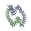 8pwlC C: citing same article ( M: map data used to model this data |
|---|---|
| Similar structure data |

-Links
 PDBj
PDBj


- Assembly
| Deposited unit | 
|
|---|---|
| 1 |
|
-Components
| #1: Protein | Mass: 99930.656 Da / Num. of mol.: 1 / Mutation: deletion 1-21 Source method: isolated from a genetically manipulated source Source: (gene. exp.) Homo sapiens (human) / Gene: HACE1, KIAA1320 / Production host:  References: UniProt: Q8IYU2, HECT-type E3 ubiquitin transferase |
|---|---|
| #2: Protein | Mass: 23769.672 Da / Num. of mol.: 1 / Mutation: Q61L Source method: isolated from a genetically manipulated source Source: (gene. exp.) Homo sapiens (human) / Gene: RAC1, TC25, MIG5 / Production host: |
| #3: Chemical | ChemComp-04E /   Mass: 185.948 Da / Num. of mol.: 1 / Source method: obtained synthetically / Formula: C2H3IO2 / Feature type: SUBJECT OF INVESTIGATION Mass: 185.948 Da / Num. of mol.: 1 / Source method: obtained synthetically / Formula: C2H3IO2 / Feature type: SUBJECT OF INVESTIGATION |
| #4: Chemical | ChemComp-GTP /   Mass: 523.180 Da / Num. of mol.: 1 / Source method: obtained synthetically / Formula: C10H16N5O14P3 / Feature type: SUBJECT OF INVESTIGATION / Comment: GTP, energy-carrying molecule*YM Mass: 523.180 Da / Num. of mol.: 1 / Source method: obtained synthetically / Formula: C10H16N5O14P3 / Feature type: SUBJECT OF INVESTIGATION / Comment: GTP, energy-carrying molecule*YM |
| Has ligand of interest | Y |
| Has protein modification | Y |
-Experimental details
-Experiment
| Experiment | Method: ELECTRON MICROSCOPY |
|---|---|
| EM experiment | Aggregation state: PARTICLE / 3D reconstruction method: single particle reconstruction |
- Sample preparation
| Component | Name: HACE1 in complex with RAC1 Q61L / Type: COMPLEX / Entity ID: #1-#2 / Source: RECOMBINANT | ||||||||||||||||
|---|---|---|---|---|---|---|---|---|---|---|---|---|---|---|---|---|---|
| Molecular weight | Experimental value: NO | ||||||||||||||||
| Source (natural) | Organism: Homo sapiens (human) | ||||||||||||||||
| Source (recombinant) | Organism: | ||||||||||||||||
| Buffer solution | pH: 8 | ||||||||||||||||
| Buffer component |
| ||||||||||||||||
| Specimen | Conc.: 0.6 mg/ml / Embedding applied: NO / Shadowing applied: NO / Staining applied: NO / Vitrification applied: YES | ||||||||||||||||
| Specimen support | Grid type: Quantifoil R1.2/1.3 | ||||||||||||||||
| Vitrification | Instrument: FEI VITROBOT MARK IV / Cryogen name: ETHANE / Humidity: 100 % / Chamber temperature: 277 K |
- Electron microscopy imaging
| Experimental equipment |  Model: Titan Krios / Image courtesy: FEI Company |
|---|---|
| Microscopy | Model: FEI TITAN KRIOS |
| Electron gun | Electron source:  FIELD EMISSION GUN / Accelerating voltage: 300 kV / Illumination mode: FLOOD BEAM FIELD EMISSION GUN / Accelerating voltage: 300 kV / Illumination mode: FLOOD BEAM |
| Electron lens | Mode: BRIGHT FIELD / Nominal magnification: 105000 X / Nominal defocus max: 3500 nm / Nominal defocus min: 800 nm |
| Image recording | Electron dose: 60 e/Å2 / Film or detector model: GATAN K3 (6k x 4k) |
- Processing
| EM software |
| |||||||||||||||||||||||||||||||||
|---|---|---|---|---|---|---|---|---|---|---|---|---|---|---|---|---|---|---|---|---|---|---|---|---|---|---|---|---|---|---|---|---|---|---|
| CTF correction | Type: PHASE FLIPPING AND AMPLITUDE CORRECTION | |||||||||||||||||||||||||||||||||
| Symmetry | Point symmetry: C1 (asymmetric) | |||||||||||||||||||||||||||||||||
| 3D reconstruction | Resolution: 4.2 Å / Resolution method: FSC 0.143 CUT-OFF / Num. of particles: 256595 / Symmetry type: POINT | |||||||||||||||||||||||||||||||||
| Atomic model building | Protocol: RIGID BODY FIT | |||||||||||||||||||||||||||||||||
| Atomic model building | Source name: AlphaFold / Type: in silico model | |||||||||||||||||||||||||||||||||
| Refine LS restraints |
|
